Iron »
PDB 6ll4-6m7e »
6ls8 »
Iron in PDB 6ls8: The Monomeric Structure of G80A/H81A/H82A Myoglobin
Protein crystallography data
The structure of The Monomeric Structure of G80A/H81A/H82A Myoglobin, PDB code: 6ls8
was solved by
S.Nagao,
A.Suda,
H.Kobayashi,
N.Shibata,
Y.Higuchi,
S.Hirota,
with X-Ray Crystallography technique. A brief refinement statistics is given in the table below:
| Resolution Low / High (Å) | 43.41 / 2.30 |
| Space group | P 1 |
| Cell size a, b, c (Å), α, β, γ (°) | 59.147, 61.490, 70.323, 78.83, 81.14, 63.12 |
| R / Rfree (%) | 21.6 / 27.4 |
Iron Binding Sites:
The binding sites of Iron atom in the The Monomeric Structure of G80A/H81A/H82A Myoglobin
(pdb code 6ls8). This binding sites where shown within
5.0 Angstroms radius around Iron atom.
In total 6 binding sites of Iron where determined in the The Monomeric Structure of G80A/H81A/H82A Myoglobin, PDB code: 6ls8:
Jump to Iron binding site number: 1; 2; 3; 4; 5; 6;
In total 6 binding sites of Iron where determined in the The Monomeric Structure of G80A/H81A/H82A Myoglobin, PDB code: 6ls8:
Jump to Iron binding site number: 1; 2; 3; 4; 5; 6;
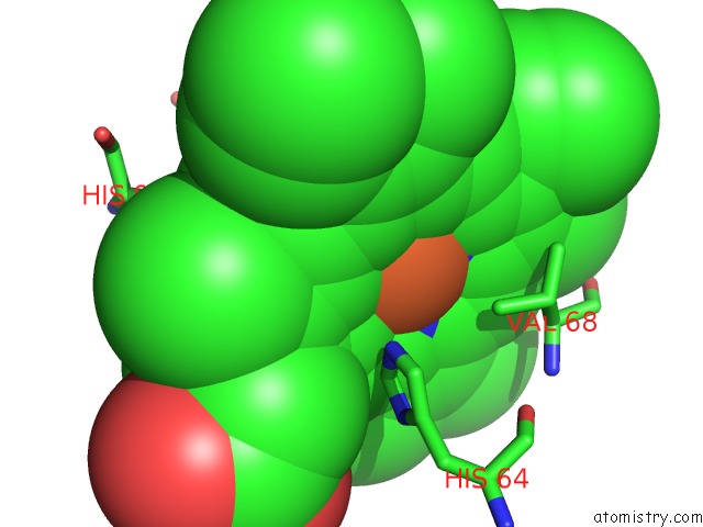
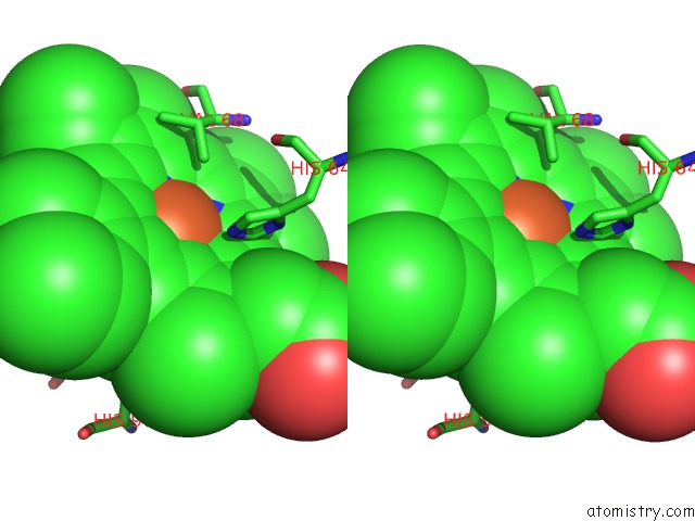
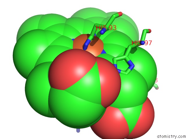
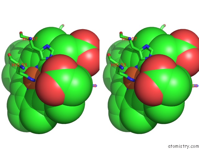
Iron binding site 1 out of 6 in 6ls8
Go back to
Iron binding site 1 out
of 6 in the The Monomeric Structure of G80A/H81A/H82A Myoglobin
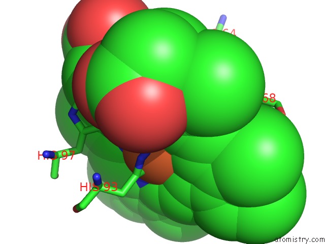
Mono view
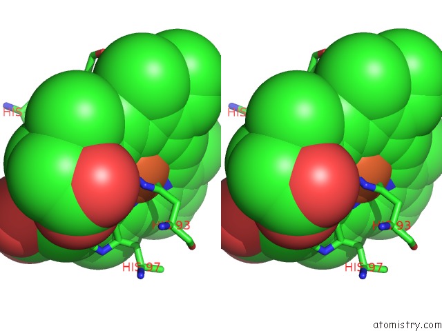
Stereo pair view
Mono view
Stereo pair view
A full contact list of Iron with other atoms in the Fe binding
site number 1 of The Monomeric Structure of G80A/H81A/H82A Myoglobin within 5.0Å range:
|
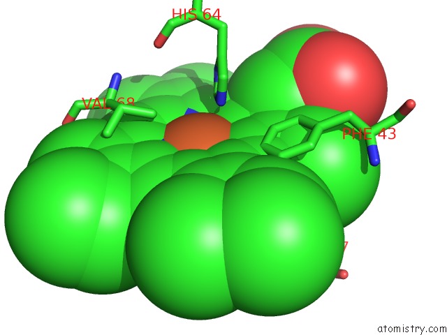
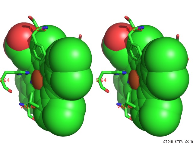
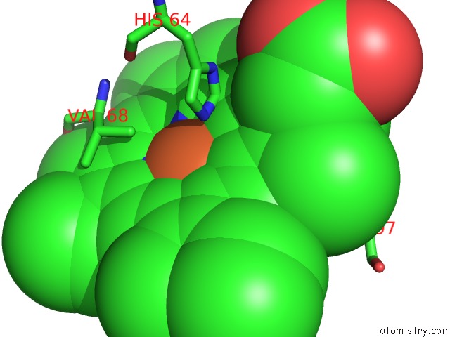
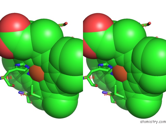
Iron binding site 2 out of 6 in 6ls8
Go back to
Iron binding site 2 out
of 6 in the The Monomeric Structure of G80A/H81A/H82A Myoglobin
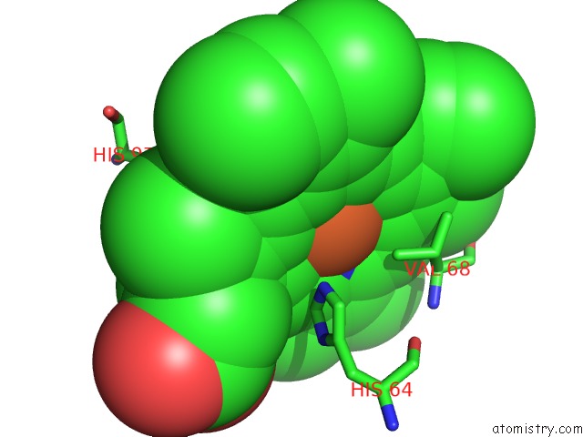
Mono view
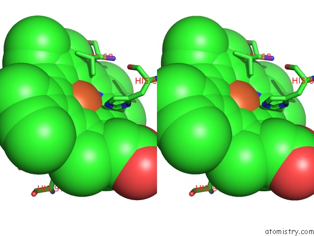
Stereo pair view
Mono view
Stereo pair view
A full contact list of Iron with other atoms in the Fe binding
site number 2 of The Monomeric Structure of G80A/H81A/H82A Myoglobin within 5.0Å range:
|
Iron binding site 3 out of 6 in 6ls8
Go back to
Iron binding site 3 out
of 6 in the The Monomeric Structure of G80A/H81A/H82A Myoglobin
Mono view
Stereo pair view
Mono view
Stereo pair view
A full contact list of Iron with other atoms in the Fe binding
site number 3 of The Monomeric Structure of G80A/H81A/H82A Myoglobin within 5.0Å range:
|
Iron binding site 4 out of 6 in 6ls8
Go back to
Iron binding site 4 out
of 6 in the The Monomeric Structure of G80A/H81A/H82A Myoglobin
Mono view
Stereo pair view
Mono view
Stereo pair view
A full contact list of Iron with other atoms in the Fe binding
site number 4 of The Monomeric Structure of G80A/H81A/H82A Myoglobin within 5.0Å range:
|
Iron binding site 5 out of 6 in 6ls8
Go back to
Iron binding site 5 out
of 6 in the The Monomeric Structure of G80A/H81A/H82A Myoglobin
Mono view
Stereo pair view
Mono view
Stereo pair view
A full contact list of Iron with other atoms in the Fe binding
site number 5 of The Monomeric Structure of G80A/H81A/H82A Myoglobin within 5.0Å range:
|
Iron binding site 6 out of 6 in 6ls8
Go back to
Iron binding site 6 out
of 6 in the The Monomeric Structure of G80A/H81A/H82A Myoglobin
Mono view
Stereo pair view
Mono view
Stereo pair view
A full contact list of Iron with other atoms in the Fe binding
site number 6 of The Monomeric Structure of G80A/H81A/H82A Myoglobin within 5.0Å range:
|
Reference:
S.Nagao,
A.Suda,
H.Kobayashi,
N.Shibata,
Y.Higuchi,
S.Hirota.
Thermodynamic Control of Domain Swapping By Modulating Helical Propensity in the Hinge Region of Myoglobin. Chem Asian J 2020.
ISSN: ESSN 1861-471X
PubMed: 32329228
DOI: 10.1002/ASIA.202000307
Page generated: Wed Aug 6 09:36:28 2025
ISSN: ESSN 1861-471X
PubMed: 32329228
DOI: 10.1002/ASIA.202000307
Last articles
Mg in 7MHAMg in 7MI6
Mg in 7MH9
Mg in 7MI3
Mg in 7MH8
Mg in 7MH4
Mg in 7MH5
Mg in 7MH1
Mg in 7MH0
Mg in 7MGZ