Iron »
PDB 6ll4-6m7e »
6m7e »
Iron in PDB 6m7e: Structure of Bovine Lactoperoxidase with Multiple Iodide Ions in the Distaline Heme Cavity.
Enzymatic activity of Structure of Bovine Lactoperoxidase with Multiple Iodide Ions in the Distaline Heme Cavity.
All present enzymatic activity of Structure of Bovine Lactoperoxidase with Multiple Iodide Ions in the Distaline Heme Cavity.:
1.11.1.7;
1.11.1.7;
Protein crystallography data
The structure of Structure of Bovine Lactoperoxidase with Multiple Iodide Ions in the Distaline Heme Cavity., PDB code: 6m7e
was solved by
A.Maurya,
V.Viswanathan,
N.Pandey,
A.K.Singh,
M.Sinha,
P.Kaur,
S.Sharma,
T.P.Singh,
with X-Ray Crystallography technique. A brief refinement statistics is given in the table below:
| Resolution Low / High (Å) | 43.95 / 2.42 |
| Space group | P 1 21 1 |
| Cell size a, b, c (Å), α, β, γ (°) | 53.810, 80.673, 74.442, 90.00, 103.07, 90.00 |
| R / Rfree (%) | 18.4 / 26.9 |
Other elements in 6m7e:
The structure of Structure of Bovine Lactoperoxidase with Multiple Iodide Ions in the Distaline Heme Cavity. also contains other interesting chemical elements:
| Iodine | (I) | 16 atoms |
| Calcium | (Ca) | 1 atom |
| Chlorine | (Cl) | 1 atom |
Iron Binding Sites:
The binding sites of Iron atom in the Structure of Bovine Lactoperoxidase with Multiple Iodide Ions in the Distaline Heme Cavity.
(pdb code 6m7e). This binding sites where shown within
5.0 Angstroms radius around Iron atom.
In total only one binding site of Iron was determined in the Structure of Bovine Lactoperoxidase with Multiple Iodide Ions in the Distaline Heme Cavity., PDB code: 6m7e:
In total only one binding site of Iron was determined in the Structure of Bovine Lactoperoxidase with Multiple Iodide Ions in the Distaline Heme Cavity., PDB code: 6m7e:
Iron binding site 1 out of 1 in 6m7e
Go back to
Iron binding site 1 out
of 1 in the Structure of Bovine Lactoperoxidase with Multiple Iodide Ions in the Distaline Heme Cavity.
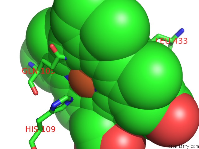
Mono view
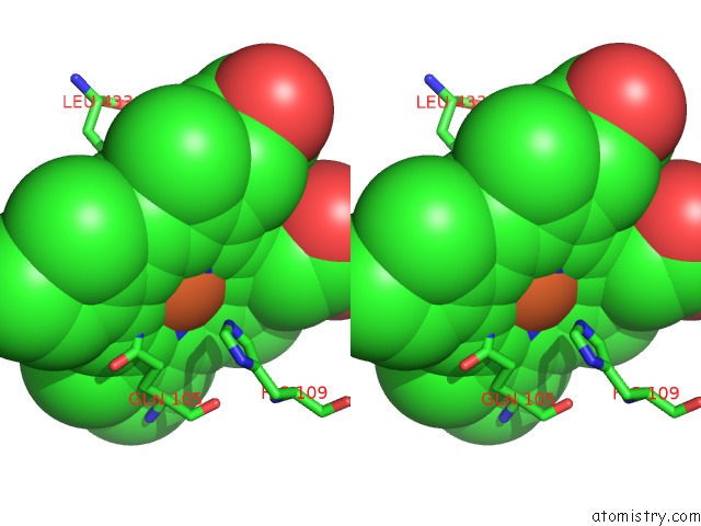
Stereo pair view
Mono view
Stereo pair view
A full contact list of Iron with other atoms in the Fe binding
site number 1 of Structure of Bovine Lactoperoxidase with Multiple Iodide Ions in the Distaline Heme Cavity. within 5.0Å range:
|
Reference:
A.Maurya,
V.Viswanathan,
N.Pandey,
A.K.Singh,
M.Sinha,
P.Kaur,
S.Sharma,
T.P.Singh.
Structure of Bovine Lactoperoxidase with Multiple Iodide Ions in the Distaline Heme Cavity. To Be Published.
Page generated: Wed Aug 6 09:51:32 2025
Last articles
Mg in 7MHAMg in 7MI6
Mg in 7MH9
Mg in 7MI3
Mg in 7MH8
Mg in 7MH4
Mg in 7MH5
Mg in 7MH1
Mg in 7MH0
Mg in 7MGZ