Iron »
PDB 6m7l-6n20 »
6mfl »
Iron in PDB 6mfl: Structure of Siderophore Binding Protein Baub Bound to A Complex Between Two Molecules of Acinetobactin and Ferric Iron.
Protein crystallography data
The structure of Structure of Siderophore Binding Protein Baub Bound to A Complex Between Two Molecules of Acinetobactin and Ferric Iron., PDB code: 6mfl
was solved by
A.M.Gulick,
D.C.Bailey,
T.A.Wencewicz,
with X-Ray Crystallography technique. A brief refinement statistics is given in the table below:
| Resolution Low / High (Å) | 56.21 / 1.90 |
| Space group | P 1 21 1 |
| Cell size a, b, c (Å), α, β, γ (°) | 37.762, 137.055, 56.208, 90.00, 90.02, 90.00 |
| R / Rfree (%) | 16.9 / 21.4 |
Iron Binding Sites:
The binding sites of Iron atom in the Structure of Siderophore Binding Protein Baub Bound to A Complex Between Two Molecules of Acinetobactin and Ferric Iron.
(pdb code 6mfl). This binding sites where shown within
5.0 Angstroms radius around Iron atom.
In total 2 binding sites of Iron where determined in the Structure of Siderophore Binding Protein Baub Bound to A Complex Between Two Molecules of Acinetobactin and Ferric Iron., PDB code: 6mfl:
Jump to Iron binding site number: 1; 2;
In total 2 binding sites of Iron where determined in the Structure of Siderophore Binding Protein Baub Bound to A Complex Between Two Molecules of Acinetobactin and Ferric Iron., PDB code: 6mfl:
Jump to Iron binding site number: 1; 2;
Iron binding site 1 out of 2 in 6mfl
Go back to
Iron binding site 1 out
of 2 in the Structure of Siderophore Binding Protein Baub Bound to A Complex Between Two Molecules of Acinetobactin and Ferric Iron.
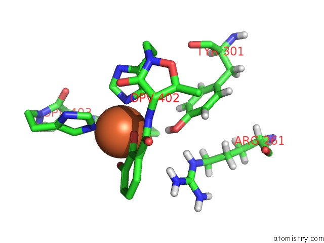
Mono view
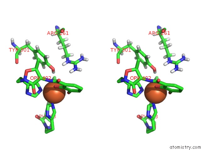
Stereo pair view
Mono view
Stereo pair view
A full contact list of Iron with other atoms in the Fe binding
site number 1 of Structure of Siderophore Binding Protein Baub Bound to A Complex Between Two Molecules of Acinetobactin and Ferric Iron. within 5.0Å range:
|
Iron binding site 2 out of 2 in 6mfl
Go back to
Iron binding site 2 out
of 2 in the Structure of Siderophore Binding Protein Baub Bound to A Complex Between Two Molecules of Acinetobactin and Ferric Iron.
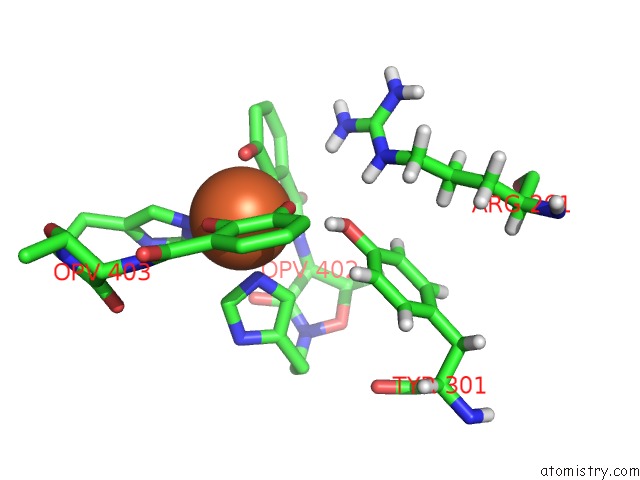
Mono view
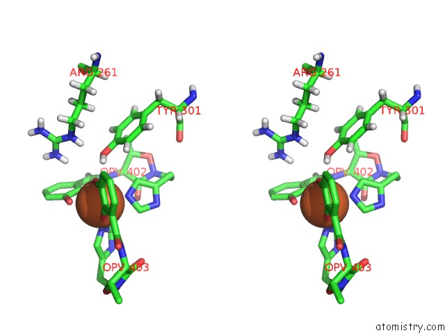
Stereo pair view
Mono view
Stereo pair view
A full contact list of Iron with other atoms in the Fe binding
site number 2 of Structure of Siderophore Binding Protein Baub Bound to A Complex Between Two Molecules of Acinetobactin and Ferric Iron. within 5.0Å range:
|
Reference:
D.C.Bailey,
T.J.Bohac,
J.A.Shapiro,
D.E.Giblin,
T.A.Wencewicz,
A.M.Gulick.
Crystal Structure of the Siderophore Binding Protein Baub Bound to An Unusual 2:1 Complex Between Acinetobactin and Ferric Iron. Biochemistry V. 57 6653 2018.
ISSN: ISSN 1520-4995
PubMed: 30406986
DOI: 10.1021/ACS.BIOCHEM.8B00986
Page generated: Wed Aug 6 09:54:33 2025
ISSN: ISSN 1520-4995
PubMed: 30406986
DOI: 10.1021/ACS.BIOCHEM.8B00986
Last articles
Mg in 6PSTMg in 6PSV
Mg in 6PSU
Mg in 6PSW
Mg in 6PGK
Mg in 6PNJ
Mg in 6PRV
Mg in 6PSS
Mg in 6PSR
Mg in 6PSQ