Iron »
PDB 6o6m-6op1 »
6on1 »
Iron in PDB 6on1: A Resting State Structure of L-Dopa Dioxygenase From Streptomyces Sclerotialus
Protein crystallography data
The structure of A Resting State Structure of L-Dopa Dioxygenase From Streptomyces Sclerotialus, PDB code: 6on1
was solved by
Y.Wang,
I.Shin,
Y.Fu,
K.Colabroy,
A.Liu,
with X-Ray Crystallography technique. A brief refinement statistics is given in the table below:
| Resolution Low / High (Å) | 33.33 / 1.98 |
| Space group | P 1 21 1 |
| Cell size a, b, c (Å), α, β, γ (°) | 99.977, 45.215, 131.153, 90.00, 105.20, 90.00 |
| R / Rfree (%) | 20.7 / 25.6 |
Iron Binding Sites:
The binding sites of Iron atom in the A Resting State Structure of L-Dopa Dioxygenase From Streptomyces Sclerotialus
(pdb code 6on1). This binding sites where shown within
5.0 Angstroms radius around Iron atom.
In total 6 binding sites of Iron where determined in the A Resting State Structure of L-Dopa Dioxygenase From Streptomyces Sclerotialus, PDB code: 6on1:
Jump to Iron binding site number: 1; 2; 3; 4; 5; 6;
In total 6 binding sites of Iron where determined in the A Resting State Structure of L-Dopa Dioxygenase From Streptomyces Sclerotialus, PDB code: 6on1:
Jump to Iron binding site number: 1; 2; 3; 4; 5; 6;
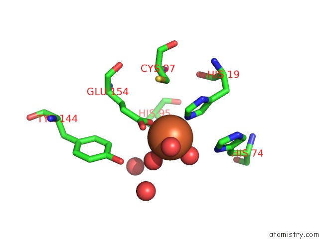
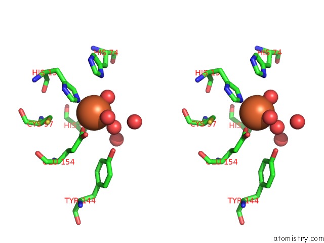
Iron binding site 1 out of 6 in 6on1
Go back to
Iron binding site 1 out
of 6 in the A Resting State Structure of L-Dopa Dioxygenase From Streptomyces Sclerotialus

Mono view

Stereo pair view
Mono view
Stereo pair view
A full contact list of Iron with other atoms in the Fe binding
site number 1 of A Resting State Structure of L-Dopa Dioxygenase From Streptomyces Sclerotialus within 5.0Å range:
|
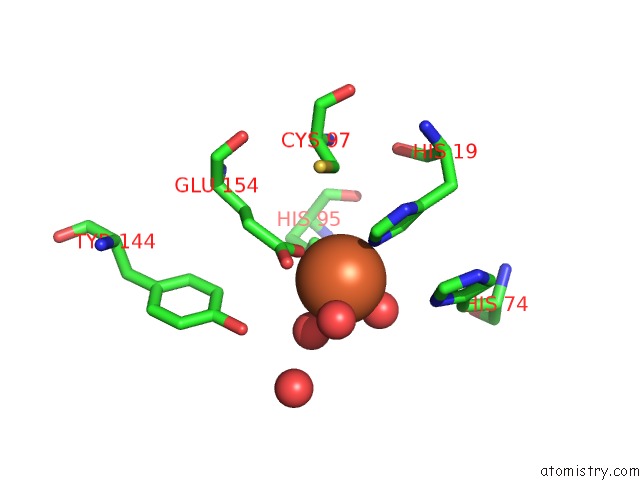
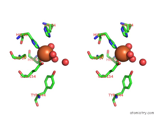
Iron binding site 2 out of 6 in 6on1
Go back to
Iron binding site 2 out
of 6 in the A Resting State Structure of L-Dopa Dioxygenase From Streptomyces Sclerotialus
Mono view

Stereo pair view
Mono view
Stereo pair view
A full contact list of Iron with other atoms in the Fe binding
site number 2 of A Resting State Structure of L-Dopa Dioxygenase From Streptomyces Sclerotialus within 5.0Å range:
|
Iron binding site 3 out of 6 in 6on1
Go back to
Iron binding site 3 out
of 6 in the A Resting State Structure of L-Dopa Dioxygenase From Streptomyces Sclerotialus

Mono view
Stereo pair view
Mono view
Stereo pair view
A full contact list of Iron with other atoms in the Fe binding
site number 3 of A Resting State Structure of L-Dopa Dioxygenase From Streptomyces Sclerotialus within 5.0Å range:
|
Iron binding site 4 out of 6 in 6on1
Go back to
Iron binding site 4 out
of 6 in the A Resting State Structure of L-Dopa Dioxygenase From Streptomyces Sclerotialus

Mono view

Stereo pair view
Mono view
Stereo pair view
A full contact list of Iron with other atoms in the Fe binding
site number 4 of A Resting State Structure of L-Dopa Dioxygenase From Streptomyces Sclerotialus within 5.0Å range:
|
Iron binding site 5 out of 6 in 6on1
Go back to
Iron binding site 5 out
of 6 in the A Resting State Structure of L-Dopa Dioxygenase From Streptomyces Sclerotialus
Mono view
Stereo pair view
Mono view
Stereo pair view
A full contact list of Iron with other atoms in the Fe binding
site number 5 of A Resting State Structure of L-Dopa Dioxygenase From Streptomyces Sclerotialus within 5.0Å range:
|
Iron binding site 6 out of 6 in 6on1
Go back to
Iron binding site 6 out
of 6 in the A Resting State Structure of L-Dopa Dioxygenase From Streptomyces Sclerotialus

Mono view

Stereo pair view
Mono view
Stereo pair view
A full contact list of Iron with other atoms in the Fe binding
site number 6 of A Resting State Structure of L-Dopa Dioxygenase From Streptomyces Sclerotialus within 5.0Å range:
|
Reference:
Y.Wang,
I.Shin,
Y.Fu,
K.L.Colabroy,
A.Liu.
Crystal Structures of L-Dopa Dioxygenase From Streptomyces Sclerotialus. Biochemistry 2019.
ISSN: ISSN 0006-2960
PubMed: 31180203
DOI: 10.1021/ACS.BIOCHEM.9B00396
Page generated: Wed Aug 7 04:29:30 2024
ISSN: ISSN 0006-2960
PubMed: 31180203
DOI: 10.1021/ACS.BIOCHEM.9B00396
Last articles
Fe in 2YXOFe in 2YRS
Fe in 2YXC
Fe in 2YNM
Fe in 2YVJ
Fe in 2YP1
Fe in 2YU2
Fe in 2YU1
Fe in 2YQB
Fe in 2YOO