Iron »
PDB 6o6m-6op1 »
6onq »
Iron in PDB 6onq: Crystal Structure of C-Type Cytochrome Xoxg From Methylobacterium Extorquens AM1
Protein crystallography data
The structure of Crystal Structure of C-Type Cytochrome Xoxg From Methylobacterium Extorquens AM1, PDB code: 6onq
was solved by
M.J.Mcbride,
E.R.Featherston,
A.K.Boal,
with X-Ray Crystallography technique. A brief refinement statistics is given in the table below:
| Resolution Low / High (Å) | 41.50 / 2.71 |
| Space group | P 62 2 2 |
| Cell size a, b, c (Å), α, β, γ (°) | 95.841, 95.841, 80.411, 90.00, 90.00, 120.00 |
| R / Rfree (%) | 19.4 / 24.8 |
Iron Binding Sites:
The binding sites of Iron atom in the Crystal Structure of C-Type Cytochrome Xoxg From Methylobacterium Extorquens AM1
(pdb code 6onq). This binding sites where shown within
5.0 Angstroms radius around Iron atom.
In total only one binding site of Iron was determined in the Crystal Structure of C-Type Cytochrome Xoxg From Methylobacterium Extorquens AM1, PDB code: 6onq:
In total only one binding site of Iron was determined in the Crystal Structure of C-Type Cytochrome Xoxg From Methylobacterium Extorquens AM1, PDB code: 6onq:
Iron binding site 1 out of 1 in 6onq
Go back to
Iron binding site 1 out
of 1 in the Crystal Structure of C-Type Cytochrome Xoxg From Methylobacterium Extorquens AM1
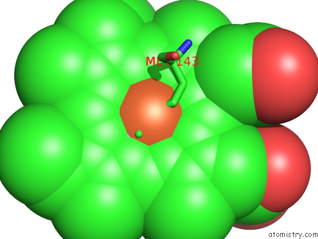
Mono view
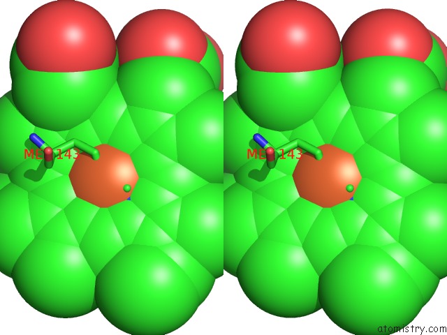
Stereo pair view
Mono view
Stereo pair view
A full contact list of Iron with other atoms in the Fe binding
site number 1 of Crystal Structure of C-Type Cytochrome Xoxg From Methylobacterium Extorquens AM1 within 5.0Å range:
|
Reference:
E.R.Featherston,
H.R.Rose,
M.J.Mcbride,
E.M.Taylor,
A.K.Boal,
J.A.Cotruvo Jr..
Biochemical and Structural Characterization of Xoxg and Xoxj and Their Roles in Lanthanide-Dependent Methanol Dehydrogenase Activity. Chembiochem V. 20 2360 2019.
ISSN: ESSN 1439-7633
PubMed: 31017712
DOI: 10.1002/CBIC.201900184
Page generated: Wed Aug 7 05:01:01 2024
ISSN: ESSN 1439-7633
PubMed: 31017712
DOI: 10.1002/CBIC.201900184
Last articles
Fe in 2YXOFe in 2YRS
Fe in 2YXC
Fe in 2YNM
Fe in 2YVJ
Fe in 2YP1
Fe in 2YU2
Fe in 2YU1
Fe in 2YQB
Fe in 2YOO