Iron »
PDB 6r2q-6rr1 »
6r4q »
Iron in PDB 6r4q: Crystal Structure of the Periplasmic Nickel-Binding Protein Nika From Escherichia Coli in Complex with Ru(Bpza)Co H2O Cl
Protein crystallography data
The structure of Crystal Structure of the Periplasmic Nickel-Binding Protein Nika From Escherichia Coli in Complex with Ru(Bpza)Co H2O Cl, PDB code: 6r4q
was solved by
C.Cavazza,
S.Menage,
with X-Ray Crystallography technique. A brief refinement statistics is given in the table below:
| Resolution Low / High (Å) | 44.52 / 1.90 |
| Space group | P 21 21 21 |
| Cell size a, b, c (Å), α, β, γ (°) | 86.629, 94.101, 124.418, 90.00, 90.00, 90.00 |
| R / Rfree (%) | 17.8 / 23.2 |
Other elements in 6r4q:
The structure of Crystal Structure of the Periplasmic Nickel-Binding Protein Nika From Escherichia Coli in Complex with Ru(Bpza)Co H2O Cl also contains other interesting chemical elements:
| Ruthenium | (Ru) | 1 atom |
| Chlorine | (Cl) | 2 atoms |
Iron Binding Sites:
The binding sites of Iron atom in the Crystal Structure of the Periplasmic Nickel-Binding Protein Nika From Escherichia Coli in Complex with Ru(Bpza)Co H2O Cl
(pdb code 6r4q). This binding sites where shown within
5.0 Angstroms radius around Iron atom.
In total only one binding site of Iron was determined in the Crystal Structure of the Periplasmic Nickel-Binding Protein Nika From Escherichia Coli in Complex with Ru(Bpza)Co H2O Cl, PDB code: 6r4q:
In total only one binding site of Iron was determined in the Crystal Structure of the Periplasmic Nickel-Binding Protein Nika From Escherichia Coli in Complex with Ru(Bpza)Co H2O Cl, PDB code: 6r4q:
Iron binding site 1 out of 1 in 6r4q
Go back to
Iron binding site 1 out
of 1 in the Crystal Structure of the Periplasmic Nickel-Binding Protein Nika From Escherichia Coli in Complex with Ru(Bpza)Co H2O Cl
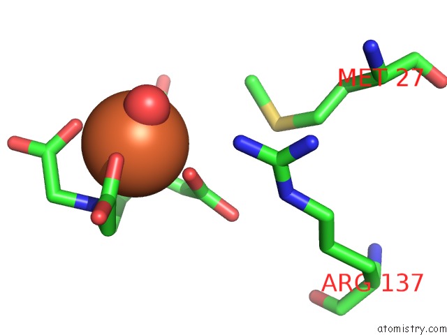
Mono view
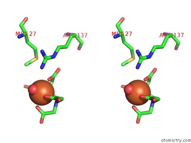
Stereo pair view
Mono view
Stereo pair view
A full contact list of Iron with other atoms in the Fe binding
site number 1 of Crystal Structure of the Periplasmic Nickel-Binding Protein Nika From Escherichia Coli in Complex with Ru(Bpza)Co H2O Cl within 5.0Å range:
|
Reference:
C.Cavazza,
S.Menage.
Crystal Structure of the Periplasmic Nickel-Binding Protein Nika From Escherichia Coli in Complex with Ru(Bpza)Co H2O Cl To Be Published.
Page generated: Wed Aug 7 08:39:55 2024
Last articles
Fe in 2YXOFe in 2YRS
Fe in 2YXC
Fe in 2YNM
Fe in 2YVJ
Fe in 2YP1
Fe in 2YU2
Fe in 2YU1
Fe in 2YQB
Fe in 2YOO