Iron »
PDB 6soy-6tmf »
6sv3 »
Iron in PDB 6sv3: Structure of Coproheme-Lmcpfc
Enzymatic activity of Structure of Coproheme-Lmcpfc
All present enzymatic activity of Structure of Coproheme-Lmcpfc:
4.99.1.1;
4.99.1.1;
Protein crystallography data
The structure of Structure of Coproheme-Lmcpfc, PDB code: 6sv3
was solved by
S.Hofbauer,
J.Helm,
K.Djinovic-Carugo,
P.G.Furtmueller,
with X-Ray Crystallography technique. A brief refinement statistics is given in the table below:
| Resolution Low / High (Å) | 36.50 / 1.64 |
| Space group | P 1 21 1 |
| Cell size a, b, c (Å), α, β, γ (°) | 37.470, 68.140, 62.910, 90.00, 103.06, 90.00 |
| R / Rfree (%) | 17.1 / 20.1 |
Iron Binding Sites:
The binding sites of Iron atom in the Structure of Coproheme-Lmcpfc
(pdb code 6sv3). This binding sites where shown within
5.0 Angstroms radius around Iron atom.
In total only one binding site of Iron was determined in the Structure of Coproheme-Lmcpfc, PDB code: 6sv3:
In total only one binding site of Iron was determined in the Structure of Coproheme-Lmcpfc, PDB code: 6sv3:
Iron binding site 1 out of 1 in 6sv3
Go back to
Iron binding site 1 out
of 1 in the Structure of Coproheme-Lmcpfc
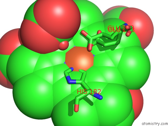
Mono view
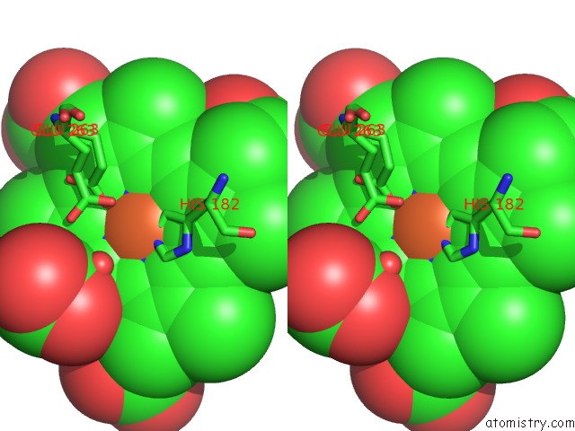
Stereo pair view
Mono view
Stereo pair view
A full contact list of Iron with other atoms in the Fe binding
site number 1 of Structure of Coproheme-Lmcpfc within 5.0Å range:
|
Reference:
S.Hofbauer,
J.Helm,
C.Obinger,
K.Djinovic-Carugo,
P.G.Furtmuller.
Crystal Structures and Calorimetry Reveal Catalytically Relevant Binding Mode of Coproporphyrin and Coproheme in Coproporphyrin Ferrochelatase. Febs J. 2019.
ISSN: ISSN 1742-464X
PubMed: 31794133
DOI: 10.1111/FEBS.15164
Page generated: Wed Aug 6 13:50:07 2025
ISSN: ISSN 1742-464X
PubMed: 31794133
DOI: 10.1111/FEBS.15164
Last articles
Mg in 4DR7Mg in 4DR6
Mg in 4DR5
Mg in 4DUX
Mg in 4DUW
Mg in 4DUV
Mg in 4DUO
Mg in 4DUG
Mg in 4DTY
Mg in 4DTW