Iron »
PDB 6soy-6tmf »
6sva »
Iron in PDB 6sva: Multicrystal Structure of Equine Haemoglobin at Room Temperature Using A Multilayer Monochromator.
Protein crystallography data
The structure of Multicrystal Structure of Equine Haemoglobin at Room Temperature Using A Multilayer Monochromator., PDB code: 6sva
was solved by
J.Sandy,
J.Sanchez-Weatherby,
H.Mikolajek,
G.Lewis,
R.Angus,
with X-Ray Crystallography technique. A brief refinement statistics is given in the table below:
| Resolution Low / High (Å) | 50.64 / 1.92 |
| Space group | C 1 2 1 |
| Cell size a, b, c (Å), α, β, γ (°) | 108.515, 62.992, 54.591, 90.00, 111.05, 90.00 |
| R / Rfree (%) | 14.1 / 17.3 |
Iron Binding Sites:
The binding sites of Iron atom in the Multicrystal Structure of Equine Haemoglobin at Room Temperature Using A Multilayer Monochromator.
(pdb code 6sva). This binding sites where shown within
5.0 Angstroms radius around Iron atom.
In total 2 binding sites of Iron where determined in the Multicrystal Structure of Equine Haemoglobin at Room Temperature Using A Multilayer Monochromator., PDB code: 6sva:
Jump to Iron binding site number: 1; 2;
In total 2 binding sites of Iron where determined in the Multicrystal Structure of Equine Haemoglobin at Room Temperature Using A Multilayer Monochromator., PDB code: 6sva:
Jump to Iron binding site number: 1; 2;
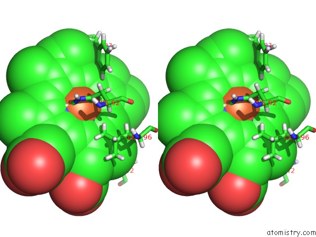
Iron binding site 1 out of 2 in 6sva
Go back to
Iron binding site 1 out
of 2 in the Multicrystal Structure of Equine Haemoglobin at Room Temperature Using A Multilayer Monochromator.

Mono view

Stereo pair view
Mono view
Stereo pair view
A full contact list of Iron with other atoms in the Fe binding
site number 1 of Multicrystal Structure of Equine Haemoglobin at Room Temperature Using A Multilayer Monochromator. within 5.0Å range:
|
Iron binding site 2 out of 2 in 6sva
Go back to
Iron binding site 2 out
of 2 in the Multicrystal Structure of Equine Haemoglobin at Room Temperature Using A Multilayer Monochromator.

Mono view
Stereo pair view
Mono view
Stereo pair view
A full contact list of Iron with other atoms in the Fe binding
site number 2 of Multicrystal Structure of Equine Haemoglobin at Room Temperature Using A Multilayer Monochromator. within 5.0Å range:
|
Reference:
J.Sandy,
J.Sanchez-Weatherby,
H.Mikolajek,
G.Lewis,
R.Angus.
Multicrystal Structure of Equine Haemoglobin at Room Temperature Using A Multilayer Monochromator. To Be Published.
Page generated: Wed Aug 6 13:50:16 2025
Last articles
Mg in 4UAYMg in 4UAS
Mg in 4UAU
Mg in 4UAT
Mg in 4UAV
Mg in 4U9U
Mg in 4UAK
Mg in 4U9I
Mg in 4U9L
Mg in 4U67