Iron »
PDB 6soy-6tmf »
6tae »
Iron in PDB 6tae: Neutron Structure of Ferric Ascorbate Peroxidase
Enzymatic activity of Neutron Structure of Ferric Ascorbate Peroxidase
All present enzymatic activity of Neutron Structure of Ferric Ascorbate Peroxidase:
1.11.1.11;
1.11.1.11;
Protein crystallography data
The structure of Neutron Structure of Ferric Ascorbate Peroxidase, PDB code: 6tae
was solved by
H.Kwon,
J.Basran,
J.M.Devos,
T.E.Schrader,
A.Ostermann,
M.P.Blakeley,
E.L.Raven,
P.C.E.Moody,
with X-Ray Crystallography technique. A brief refinement statistics is given in the table below:
| Resolution Low / High (Å) | N/A / 1.90 |
| Space group | P 42 21 2 |
| Cell size a, b, c (Å), α, β, γ (°) | 82.904, 82.904, 75.868, 90.00, 90.00, 90.00 |
| R / Rfree (%) | 27.6 / 30.8 |
Iron Binding Sites:
The binding sites of Iron atom in the Neutron Structure of Ferric Ascorbate Peroxidase
(pdb code 6tae). This binding sites where shown within
5.0 Angstroms radius around Iron atom.
In total only one binding site of Iron was determined in the Neutron Structure of Ferric Ascorbate Peroxidase, PDB code: 6tae:
In total only one binding site of Iron was determined in the Neutron Structure of Ferric Ascorbate Peroxidase, PDB code: 6tae:
Iron binding site 1 out of 1 in 6tae
Go back to
Iron binding site 1 out
of 1 in the Neutron Structure of Ferric Ascorbate Peroxidase

Mono view
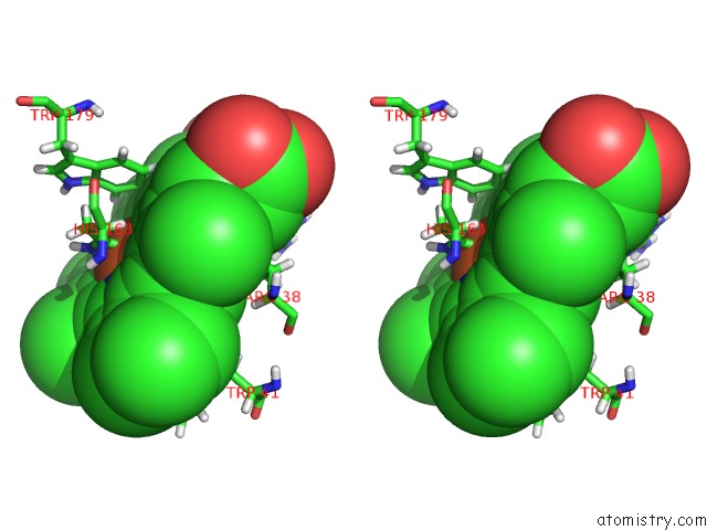
Stereo pair view
Mono view
Stereo pair view
A full contact list of Iron with other atoms in the Fe binding
site number 1 of Neutron Structure of Ferric Ascorbate Peroxidase within 5.0Å range:
|
Reference:
H.Kwon,
J.Basran,
J.M.Devos,
R.Suardiaz,
M.W.Van Der Kamp,
A.J.Mulholland,
T.E.Schrader,
A.Ostermann,
M.P.Blakeley,
P.C.E.Moody,
E.L.Raven.
Visualizing the Protons in A Metalloenzyme Electron Proton Transfer Pathway. Proc.Natl.Acad.Sci.Usa 2020.
ISSN: ESSN 1091-6490
PubMed: 32152099
DOI: 10.1073/PNAS.1918936117
Page generated: Wed Aug 6 13:57:12 2025
ISSN: ESSN 1091-6490
PubMed: 32152099
DOI: 10.1073/PNAS.1918936117
Last articles
Mg in 4Q66Mg in 4Q8B
Mg in 4Q7F
Mg in 4Q5S
Mg in 4Q6X
Mg in 4Q4Z
Mg in 4Q5H
Mg in 4Q4C
Mg in 4Q4D
Mg in 4Q57