Iron »
PDB 6vlt-6wnc »
6w0s »
Iron in PDB 6w0s: Crystal Structure of Substrate Free Cytochrome P450 NASF5053 From Streptomyces Sp. Nrrl F-5053
Protein crystallography data
The structure of Crystal Structure of Substrate Free Cytochrome P450 NASF5053 From Streptomyces Sp. Nrrl F-5053, PDB code: 6w0s
was solved by
Z.Luo,
X.Jia,
C.Sun,
X.Qu,
B.Kobe,
with X-Ray Crystallography technique. A brief refinement statistics is given in the table below:
| Resolution Low / High (Å) | 43.06 / 1.70 |
| Space group | P 1 21 1 |
| Cell size a, b, c (Å), α, β, γ (°) | 42.208, 91.325, 98.275, 90.00, 96.41, 90.00 |
| R / Rfree (%) | 18.6 / 22 |
Other elements in 6w0s:
The structure of Crystal Structure of Substrate Free Cytochrome P450 NASF5053 From Streptomyces Sp. Nrrl F-5053 also contains other interesting chemical elements:
| Bromine | (Br) | 4 atoms |
| Chlorine | (Cl) | 6 atoms |
| Sodium | (Na) | 2 atoms |
Iron Binding Sites:
The binding sites of Iron atom in the Crystal Structure of Substrate Free Cytochrome P450 NASF5053 From Streptomyces Sp. Nrrl F-5053
(pdb code 6w0s). This binding sites where shown within
5.0 Angstroms radius around Iron atom.
In total 2 binding sites of Iron where determined in the Crystal Structure of Substrate Free Cytochrome P450 NASF5053 From Streptomyces Sp. Nrrl F-5053, PDB code: 6w0s:
Jump to Iron binding site number: 1; 2;
In total 2 binding sites of Iron where determined in the Crystal Structure of Substrate Free Cytochrome P450 NASF5053 From Streptomyces Sp. Nrrl F-5053, PDB code: 6w0s:
Jump to Iron binding site number: 1; 2;
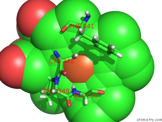
Iron binding site 1 out of 2 in 6w0s
Go back to
Iron binding site 1 out
of 2 in the Crystal Structure of Substrate Free Cytochrome P450 NASF5053 From Streptomyces Sp. Nrrl F-5053
Mono view

Stereo pair view
Mono view
Stereo pair view
A full contact list of Iron with other atoms in the Fe binding
site number 1 of Crystal Structure of Substrate Free Cytochrome P450 NASF5053 From Streptomyces Sp. Nrrl F-5053 within 5.0Å range:
|
Iron binding site 2 out of 2 in 6w0s
Go back to
Iron binding site 2 out
of 2 in the Crystal Structure of Substrate Free Cytochrome P450 NASF5053 From Streptomyces Sp. Nrrl F-5053

Mono view

Stereo pair view
Mono view
Stereo pair view
A full contact list of Iron with other atoms in the Fe binding
site number 2 of Crystal Structure of Substrate Free Cytochrome P450 NASF5053 From Streptomyces Sp. Nrrl F-5053 within 5.0Å range:
|
Reference:
C.Sun,
Z.Luo,
W.Zhang,
Z.Deng,
M.Mobli,
B.Kobe,
X.Jia,
X.Qu.
Molecular Basis of Regio- and Stereo-Specificity in Biosynthesis of Bacterial Heterodimeric Diketopiperazines To Be Published.
Page generated: Wed Aug 6 15:23:20 2025
Last articles
Mg in 3C8VMg in 3C84
Mg in 3C7T
Mg in 3C5I
Mg in 3C7N
Mg in 3C7K
Mg in 3C6H
Mg in 3C6A
Mg in 3C5C
Mg in 3C5H