Iron »
PDB 6xai-6xyv »
6xma »
Iron in PDB 6xma: Crystal Structure of Iron-Bound LSD4 From Sphingobium Sp. Strain Syk-6
Protein crystallography data
The structure of Crystal Structure of Iron-Bound LSD4 From Sphingobium Sp. Strain Syk-6, PDB code: 6xma
was solved by
E.Kuatsjah,
A.C.Chan,
R.Katahira,
G.T.Beckham,
M.E.Murphy,
L.D.Eltis,
with X-Ray Crystallography technique. A brief refinement statistics is given in the table below:
| Resolution Low / High (Å) | 29.79 / 1.45 |
| Space group | I 2 2 2 |
| Cell size a, b, c (Å), α, β, γ (°) | 86.03, 112.527, 115.384, 90, 90, 90 |
| R / Rfree (%) | 14.9 / 17.1 |
Iron Binding Sites:
The binding sites of Iron atom in the Crystal Structure of Iron-Bound LSD4 From Sphingobium Sp. Strain Syk-6
(pdb code 6xma). This binding sites where shown within
5.0 Angstroms radius around Iron atom.
In total only one binding site of Iron was determined in the Crystal Structure of Iron-Bound LSD4 From Sphingobium Sp. Strain Syk-6, PDB code: 6xma:
In total only one binding site of Iron was determined in the Crystal Structure of Iron-Bound LSD4 From Sphingobium Sp. Strain Syk-6, PDB code: 6xma:
Iron binding site 1 out of 1 in 6xma
Go back to
Iron binding site 1 out
of 1 in the Crystal Structure of Iron-Bound LSD4 From Sphingobium Sp. Strain Syk-6

Mono view
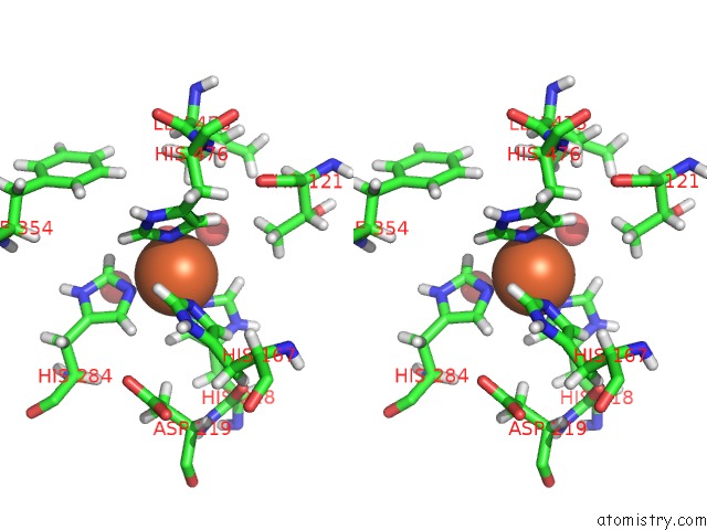
Stereo pair view
Mono view
Stereo pair view
A full contact list of Iron with other atoms in the Fe binding
site number 1 of Crystal Structure of Iron-Bound LSD4 From Sphingobium Sp. Strain Syk-6 within 5.0Å range:
|
Reference:
E.Kuatsjah,
A.C.K.Chan,
R.Katahira,
S.J.Haugen,
G.T.Beckham,
M.E.Murphy,
L.D.Eltis.
Structural and Functional Analysis of Lignostilbene Dioxygenases From Sphingobium Sp. Syk-6 J.Biol.Chem. 2021.
ISSN: ESSN 1083-351X
Page generated: Wed Aug 7 15:35:32 2024
ISSN: ESSN 1083-351X
Last articles
Br in 1XU3Br in 1XQ0
Br in 1XOK
Br in 1VP6
Br in 1XL5
Br in 1X84
Br in 1WY0
Br in 1WTP
Br in 1UNJ
Br in 1WCT