Iron »
PDB 7ai9-7bhb »
7aik »
Iron in PDB 7aik: Ribonucleotide Reductase R2 Protein From Aquifex Aeolicus
Enzymatic activity of Ribonucleotide Reductase R2 Protein From Aquifex Aeolicus
All present enzymatic activity of Ribonucleotide Reductase R2 Protein From Aquifex Aeolicus:
1.17.4.1;
1.17.4.1;
Protein crystallography data
The structure of Ribonucleotide Reductase R2 Protein From Aquifex Aeolicus, PDB code: 7aik
was solved by
D.Rehling,
E.R.Scaletti,
P.Stenmark,
with X-Ray Crystallography technique. A brief refinement statistics is given in the table below:
| Resolution Low / High (Å) | 47.37 / 2.10 |
| Space group | P 43 21 2 |
| Cell size a, b, c (Å), α, β, γ (°) | 69.42, 69.42, 177.973, 90, 90, 90 |
| R / Rfree (%) | 21.7 / 26.7 |
Iron Binding Sites:
The binding sites of Iron atom in the Ribonucleotide Reductase R2 Protein From Aquifex Aeolicus
(pdb code 7aik). This binding sites where shown within
5.0 Angstroms radius around Iron atom.
In total 2 binding sites of Iron where determined in the Ribonucleotide Reductase R2 Protein From Aquifex Aeolicus, PDB code: 7aik:
Jump to Iron binding site number: 1; 2;
In total 2 binding sites of Iron where determined in the Ribonucleotide Reductase R2 Protein From Aquifex Aeolicus, PDB code: 7aik:
Jump to Iron binding site number: 1; 2;
Iron binding site 1 out of 2 in 7aik
Go back to
Iron binding site 1 out
of 2 in the Ribonucleotide Reductase R2 Protein From Aquifex Aeolicus
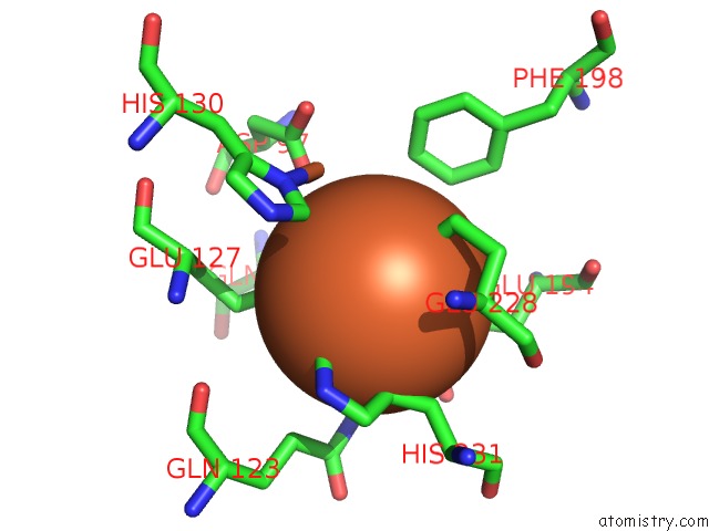
Mono view
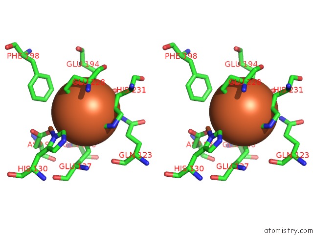
Stereo pair view
Mono view
Stereo pair view
A full contact list of Iron with other atoms in the Fe binding
site number 1 of Ribonucleotide Reductase R2 Protein From Aquifex Aeolicus within 5.0Å range:
|
Iron binding site 2 out of 2 in 7aik
Go back to
Iron binding site 2 out
of 2 in the Ribonucleotide Reductase R2 Protein From Aquifex Aeolicus
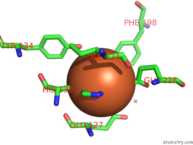
Mono view
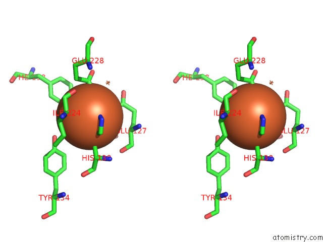
Stereo pair view
Mono view
Stereo pair view
A full contact list of Iron with other atoms in the Fe binding
site number 2 of Ribonucleotide Reductase R2 Protein From Aquifex Aeolicus within 5.0Å range:
|
Reference:
D.Rehling,
E.R.Scaletti,
P.Stenmark.
Ribonucleotide Reductase R2 Protein From Aquifex Aeolicus To Be Published.
Page generated: Wed Aug 6 19:18:39 2025
Last articles
Zn in 1VGXZn in 1VFY
Zn in 1VH2
Zn in 1VFN
Zn in 1VEZ
Zn in 1VEY
Zn in 1VEV
Zn in 1VFL
Zn in 1VF2
Zn in 1VDD