Iron »
PDB 7ck8-7czl »
7cy5 »
Iron in PDB 7cy5: Crystal Structure of CMD1 in Complex with Vitamin C
Protein crystallography data
The structure of Crystal Structure of CMD1 in Complex with Vitamin C, PDB code: 7cy5
was solved by
W.Li,
T.Zhang,
M.Sun,
J.Ding,
with X-Ray Crystallography technique. A brief refinement statistics is given in the table below:
| Resolution Low / High (Å) | 48.38 / 2.20 |
| Space group | C 1 2 1 |
| Cell size a, b, c (Å), α, β, γ (°) | 153.846, 126.418, 64.217, 90, 102.27, 90 |
| R / Rfree (%) | 18.1 / 22.1 |
Iron Binding Sites:
The binding sites of Iron atom in the Crystal Structure of CMD1 in Complex with Vitamin C
(pdb code 7cy5). This binding sites where shown within
5.0 Angstroms radius around Iron atom.
In total only one binding site of Iron was determined in the Crystal Structure of CMD1 in Complex with Vitamin C, PDB code: 7cy5:
In total only one binding site of Iron was determined in the Crystal Structure of CMD1 in Complex with Vitamin C, PDB code: 7cy5:
Iron binding site 1 out of 1 in 7cy5
Go back to
Iron binding site 1 out
of 1 in the Crystal Structure of CMD1 in Complex with Vitamin C
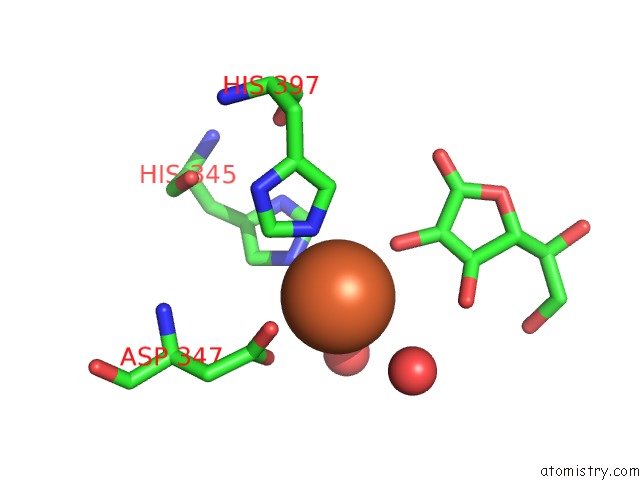
Mono view
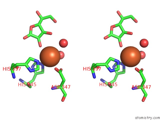
Stereo pair view
Mono view
Stereo pair view
A full contact list of Iron with other atoms in the Fe binding
site number 1 of Crystal Structure of CMD1 in Complex with Vitamin C within 5.0Å range:
|
Reference:
W.Li,
T.Zhang,
M.Sun,
Y.Shi,
X.Zhang,
G.Xu,
J.Ding.
Crystal Structures of Algal Tet Homologue CMD1 Provide Insight Into the Molecular Mechanism For Vitamin C-Derived C5-Glyceryl-Methylcytosine Dna Modification To Be Published.
Page generated: Wed Aug 6 21:27:51 2025
Last articles
Zn in 1SHNZn in 1SLX
Zn in 1SFO
Zn in 1SKU
Zn in 1SHQ
Zn in 1SHW
Zn in 1SGF
Zn in 1SG0
Zn in 1SG6
Zn in 1SED