Iron »
PDB 7d0j-7dgk »
7d2i »
Iron in PDB 7d2i: Crystal Structure of Ixodes Scapularis Glutaminyl Cyclase with A Fe Ion Bound to the Active Site
Enzymatic activity of Crystal Structure of Ixodes Scapularis Glutaminyl Cyclase with A Fe Ion Bound to the Active Site
All present enzymatic activity of Crystal Structure of Ixodes Scapularis Glutaminyl Cyclase with A Fe Ion Bound to the Active Site:
2.3.2.5;
2.3.2.5;
Protein crystallography data
The structure of Crystal Structure of Ixodes Scapularis Glutaminyl Cyclase with A Fe Ion Bound to the Active Site, PDB code: 7d2i
was solved by
K.-F.Huang,
J.-S.Huang,
M.-L.Wu,
W.-L.Hsieh,
A.H.-J.Wang,
with X-Ray Crystallography technique. A brief refinement statistics is given in the table below:
| Resolution Low / High (Å) | 26.80 / 1.85 |
| Space group | P 21 21 21 |
| Cell size a, b, c (Å), α, β, γ (°) | 55.629, 71.478, 80.926, 90, 90, 90 |
| R / Rfree (%) | 17.8 / 22.8 |
Iron Binding Sites:
The binding sites of Iron atom in the Crystal Structure of Ixodes Scapularis Glutaminyl Cyclase with A Fe Ion Bound to the Active Site
(pdb code 7d2i). This binding sites where shown within
5.0 Angstroms radius around Iron atom.
In total only one binding site of Iron was determined in the Crystal Structure of Ixodes Scapularis Glutaminyl Cyclase with A Fe Ion Bound to the Active Site, PDB code: 7d2i:
In total only one binding site of Iron was determined in the Crystal Structure of Ixodes Scapularis Glutaminyl Cyclase with A Fe Ion Bound to the Active Site, PDB code: 7d2i:
Iron binding site 1 out of 1 in 7d2i
Go back to
Iron binding site 1 out
of 1 in the Crystal Structure of Ixodes Scapularis Glutaminyl Cyclase with A Fe Ion Bound to the Active Site
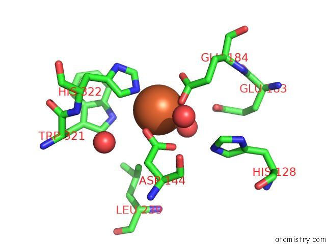
Mono view
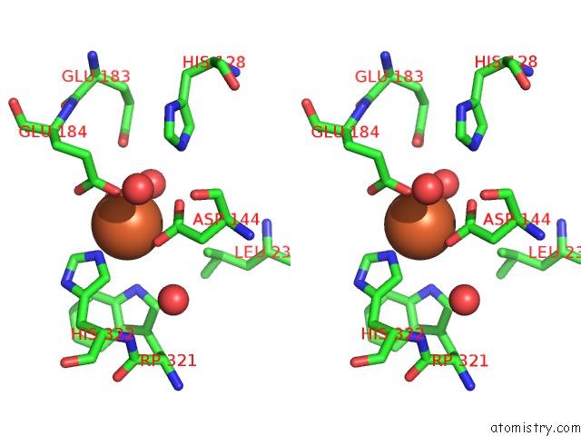
Stereo pair view
Mono view
Stereo pair view
A full contact list of Iron with other atoms in the Fe binding
site number 1 of Crystal Structure of Ixodes Scapularis Glutaminyl Cyclase with A Fe Ion Bound to the Active Site within 5.0Å range:
|
Reference:
K.F.Huang,
J.S.Huang,
M.L.Wu,
W.L.Hsieh,
K.C.Hsu,
H.L.Hsu,
T.P.Ko,
A.H-J Wang.
A Unique Carboxylic-Acid Hydrogen-Bond Network (Cahbn) Confers Glutaminyl Cyclase Activity on M28 Family Enzymes. J.Mol.Biol. 66960 2021.
ISSN: ESSN 1089-8638
PubMed: 33774034
DOI: 10.1016/J.JMB.2021.166960
Page generated: Wed Aug 6 21:31:01 2025
ISSN: ESSN 1089-8638
PubMed: 33774034
DOI: 10.1016/J.JMB.2021.166960
Last articles
Zn in 8WZYZn in 8WYX
Zn in 8WYY
Zn in 8WYU
Zn in 8WXZ
Zn in 8WYT
Zn in 8WXW
Zn in 8WUG
Zn in 8WQH
Zn in 8WTC