Iron »
PDB 7k5h-7kvp »
7kqu »
Iron in PDB 7kqu: A 1.58-A Resolution Crystal Structure of Ferric-Hydroperoxo Intermediate of L-Tyrosine Hydroxylase in Complex with 3-Fluoro-L- Tyrosine
Protein crystallography data
The structure of A 1.58-A Resolution Crystal Structure of Ferric-Hydroperoxo Intermediate of L-Tyrosine Hydroxylase in Complex with 3-Fluoro-L- Tyrosine, PDB code: 7kqu
was solved by
Y.Wang,
I.Davis,
A.Liu,
with X-Ray Crystallography technique. A brief refinement statistics is given in the table below:
| Resolution Low / High (Å) | 45.22 / 1.58 |
| Space group | P 1 21 1 |
| Cell size a, b, c (Å), α, β, γ (°) | 47.523, 129.329, 48.38, 90, 94.03, 90 |
| R / Rfree (%) | 15.7 / 18.9 |
Other elements in 7kqu:
The structure of A 1.58-A Resolution Crystal Structure of Ferric-Hydroperoxo Intermediate of L-Tyrosine Hydroxylase in Complex with 3-Fluoro-L- Tyrosine also contains other interesting chemical elements:
| Fluorine | (F) | 4 atoms |
Iron Binding Sites:
The binding sites of Iron atom in the A 1.58-A Resolution Crystal Structure of Ferric-Hydroperoxo Intermediate of L-Tyrosine Hydroxylase in Complex with 3-Fluoro-L- Tyrosine
(pdb code 7kqu). This binding sites where shown within
5.0 Angstroms radius around Iron atom.
In total 2 binding sites of Iron where determined in the A 1.58-A Resolution Crystal Structure of Ferric-Hydroperoxo Intermediate of L-Tyrosine Hydroxylase in Complex with 3-Fluoro-L- Tyrosine, PDB code: 7kqu:
Jump to Iron binding site number: 1; 2;
In total 2 binding sites of Iron where determined in the A 1.58-A Resolution Crystal Structure of Ferric-Hydroperoxo Intermediate of L-Tyrosine Hydroxylase in Complex with 3-Fluoro-L- Tyrosine, PDB code: 7kqu:
Jump to Iron binding site number: 1; 2;
Iron binding site 1 out of 2 in 7kqu
Go back to
Iron binding site 1 out
of 2 in the A 1.58-A Resolution Crystal Structure of Ferric-Hydroperoxo Intermediate of L-Tyrosine Hydroxylase in Complex with 3-Fluoro-L- Tyrosine
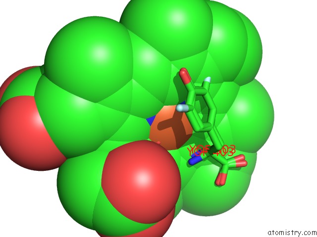
Mono view
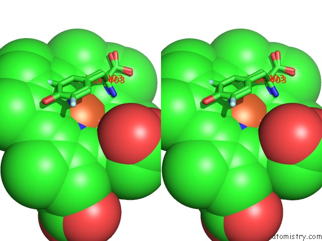
Stereo pair view
Mono view
Stereo pair view
A full contact list of Iron with other atoms in the Fe binding
site number 1 of A 1.58-A Resolution Crystal Structure of Ferric-Hydroperoxo Intermediate of L-Tyrosine Hydroxylase in Complex with 3-Fluoro-L- Tyrosine within 5.0Å range:
|
Iron binding site 2 out of 2 in 7kqu
Go back to
Iron binding site 2 out
of 2 in the A 1.58-A Resolution Crystal Structure of Ferric-Hydroperoxo Intermediate of L-Tyrosine Hydroxylase in Complex with 3-Fluoro-L- Tyrosine
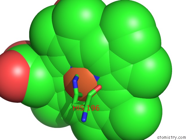
Mono view
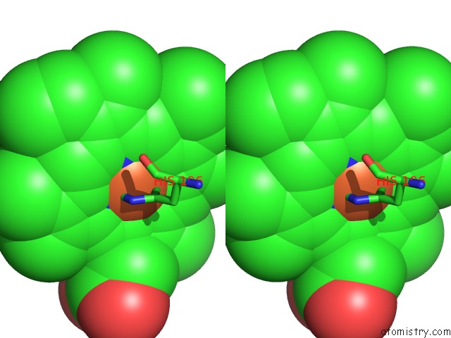
Stereo pair view
Mono view
Stereo pair view
A full contact list of Iron with other atoms in the Fe binding
site number 2 of A 1.58-A Resolution Crystal Structure of Ferric-Hydroperoxo Intermediate of L-Tyrosine Hydroxylase in Complex with 3-Fluoro-L- Tyrosine within 5.0Å range:
|
Reference:
Y.Wang,
I.Davis,
I.Shin,
H.Xu,
A.Liu.
Molecular Rationale For Partitioning Between C-H and C-F Bond Activation in Heme-Dependent Tyrosine Hydroxylase. J.Am.Chem.Soc. 2021.
ISSN: ESSN 1520-5126
PubMed: 33734681
DOI: 10.1021/JACS.1C00175
Page generated: Wed Aug 6 22:51:40 2025
ISSN: ESSN 1520-5126
PubMed: 33734681
DOI: 10.1021/JACS.1C00175
Last articles
K in 6WLVK in 6WMG
K in 6WMF
K in 6WME
K in 6WH5
K in 6WIC
K in 6WFL
K in 6WDY
K in 6WDX
K in 6WE6