Iron »
PDB 7lw8-7mh9 »
7lxl »
Iron in PDB 7lxl: Crystal Structure of Human CYP3A4 Bound to the Testosterone Dimer
Enzymatic activity of Crystal Structure of Human CYP3A4 Bound to the Testosterone Dimer
All present enzymatic activity of Crystal Structure of Human CYP3A4 Bound to the Testosterone Dimer:
1.14.14.1; 1.14.14.55; 1.14.14.56; 1.14.14.73;
1.14.14.1; 1.14.14.55; 1.14.14.56; 1.14.14.73;
Protein crystallography data
The structure of Crystal Structure of Human CYP3A4 Bound to the Testosterone Dimer, PDB code: 7lxl
was solved by
I.F.Sevrioukova,
with X-Ray Crystallography technique. A brief refinement statistics is given in the table below:
| Resolution Low / High (Å) | 40.38 / 2.75 |
| Space group | I 2 2 2 |
| Cell size a, b, c (Å), α, β, γ (°) | 77.869, 101.67, 127.7, 90, 90, 90 |
| R / Rfree (%) | 24.4 / 28.8 |
Iron Binding Sites:
The binding sites of Iron atom in the Crystal Structure of Human CYP3A4 Bound to the Testosterone Dimer
(pdb code 7lxl). This binding sites where shown within
5.0 Angstroms radius around Iron atom.
In total only one binding site of Iron was determined in the Crystal Structure of Human CYP3A4 Bound to the Testosterone Dimer, PDB code: 7lxl:
In total only one binding site of Iron was determined in the Crystal Structure of Human CYP3A4 Bound to the Testosterone Dimer, PDB code: 7lxl:
Iron binding site 1 out of 1 in 7lxl
Go back to
Iron binding site 1 out
of 1 in the Crystal Structure of Human CYP3A4 Bound to the Testosterone Dimer

Mono view
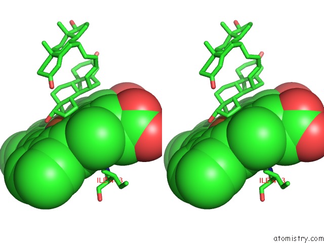
Stereo pair view
Mono view
Stereo pair view
A full contact list of Iron with other atoms in the Fe binding
site number 1 of Crystal Structure of Human CYP3A4 Bound to the Testosterone Dimer within 5.0Å range:
|
Reference:
A.Paquin,
Y.Oufqir,
I.F.Sevrioukova,
C.Reyes-Moreno,
G.Berube.
Innovative C2-Symmetric Testosterone and Androstenedione Dimers: Design, Synthesis, Biological Evaluation on Prostate Cancer Cell Lines and Binding Study to Recombinant CYP3A4 Eur.J.Med.Chem. 2021.
ISSN: ISSN 0223-5234
Page generated: Wed Aug 6 23:25:23 2025
ISSN: ISSN 0223-5234
Last articles
Mn in 9LJUMn in 9LJW
Mn in 9LJS
Mn in 9LJR
Mn in 9LJT
Mn in 9LJV
Mg in 9UA2
Mg in 9R96
Mg in 9VM1
Mg in 9P01