Iron »
PDB 7mha-7ni3 »
7n7i »
Iron in PDB 7n7i: X-Ray Crystal Structure of Viperin-Like Enzyme From Trichoderma Virens
Protein crystallography data
The structure of X-Ray Crystal Structure of Viperin-Like Enzyme From Trichoderma Virens, PDB code: 7n7i
was solved by
T.L.Grove,
S.C.Almo,
J.B.Bonanno,
J.C.Lachowicz,
A.G.Gizzi,
with X-Ray Crystallography technique. A brief refinement statistics is given in the table below:
| Resolution Low / High (Å) | 28.14 / 3.19 |
| Space group | P 31 |
| Cell size a, b, c (Å), α, β, γ (°) | 85.943, 85.943, 111.718, 90, 90, 120 |
| R / Rfree (%) | 22.6 / 26 |
Iron Binding Sites:
Pages:
>>> Page 1 <<< Page 2, Binding sites: 11 - 12;Binding sites:
The binding sites of Iron atom in the X-Ray Crystal Structure of Viperin-Like Enzyme From Trichoderma Virens (pdb code 7n7i). This binding sites where shown within 5.0 Angstroms radius around Iron atom.In total 12 binding sites of Iron where determined in the X-Ray Crystal Structure of Viperin-Like Enzyme From Trichoderma Virens, PDB code: 7n7i:
Jump to Iron binding site number: 1; 2; 3; 4; 5; 6; 7; 8; 9; 10;
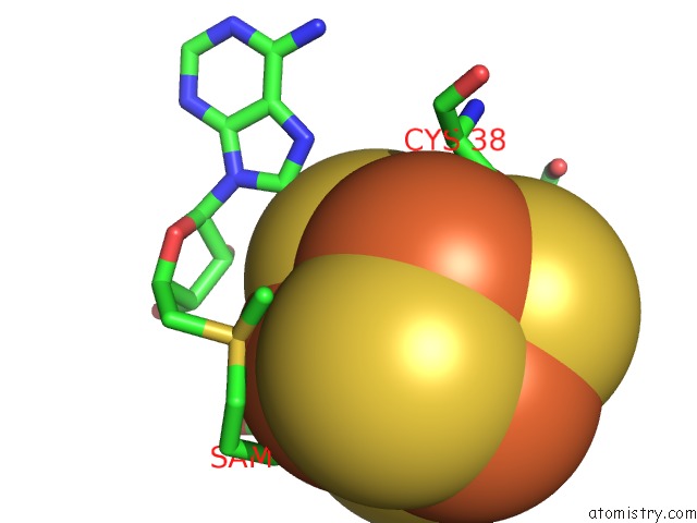
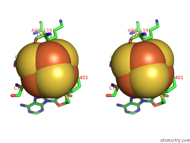
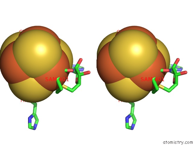
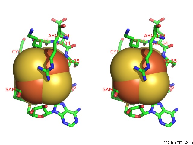
Iron binding site 1 out of 12 in 7n7i
Go back to
Iron binding site 1 out
of 12 in the X-Ray Crystal Structure of Viperin-Like Enzyme From Trichoderma Virens
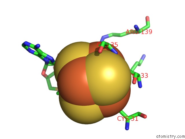
Mono view
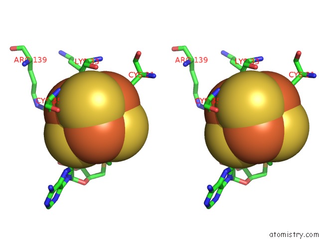
Stereo pair view
Mono view
Stereo pair view
A full contact list of Iron with other atoms in the Fe binding
site number 1 of X-Ray Crystal Structure of Viperin-Like Enzyme From Trichoderma Virens within 5.0Å range:
|
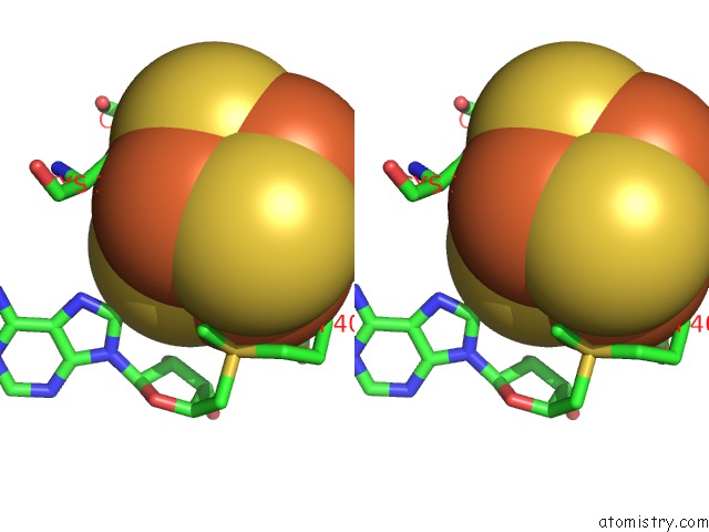
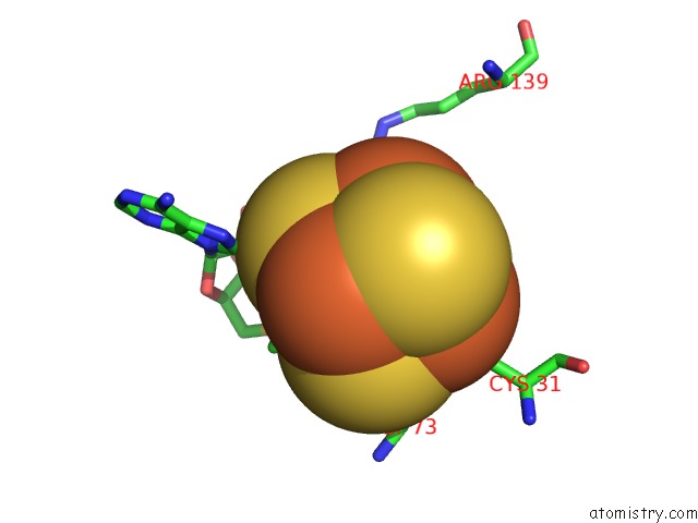
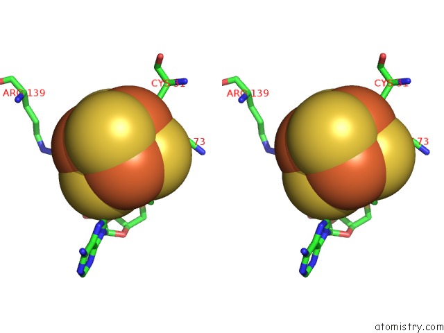
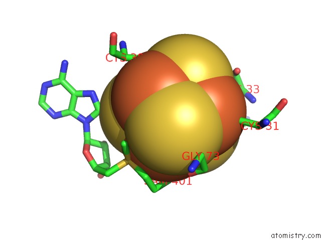
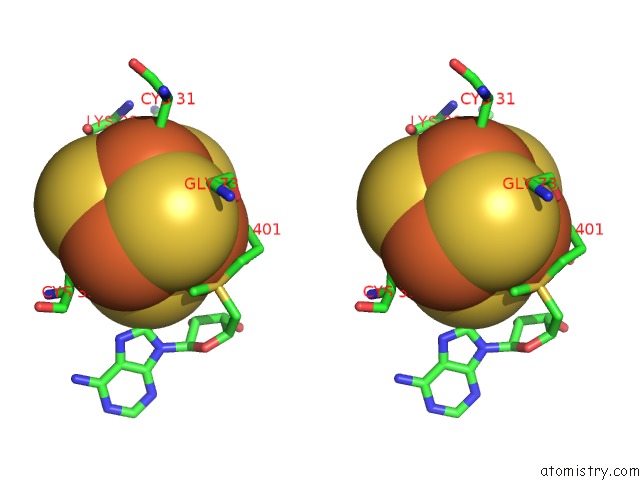
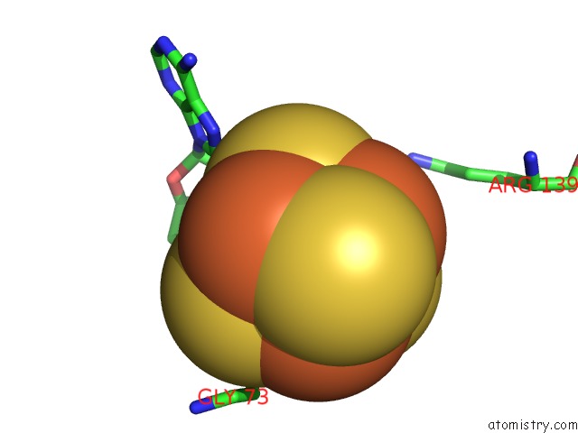
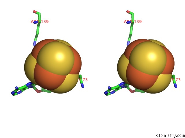
Iron binding site 2 out of 12 in 7n7i
Go back to
Iron binding site 2 out
of 12 in the X-Ray Crystal Structure of Viperin-Like Enzyme From Trichoderma Virens
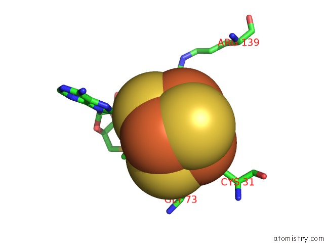
Mono view
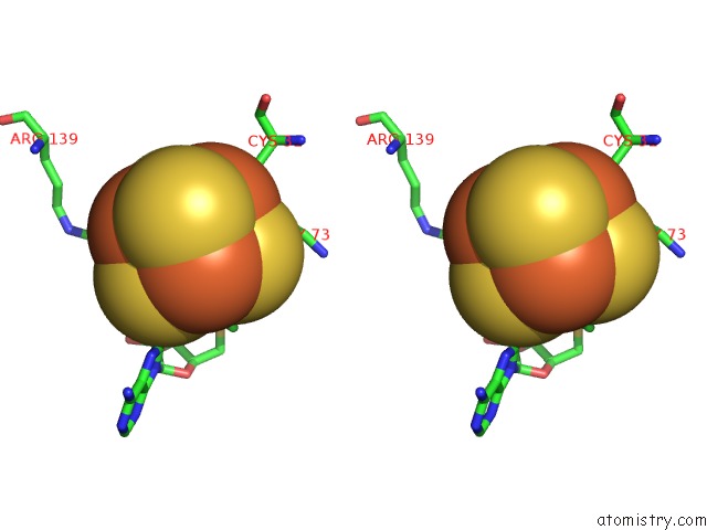
Stereo pair view
Mono view
Stereo pair view
A full contact list of Iron with other atoms in the Fe binding
site number 2 of X-Ray Crystal Structure of Viperin-Like Enzyme From Trichoderma Virens within 5.0Å range:
|
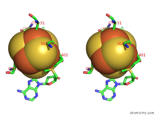
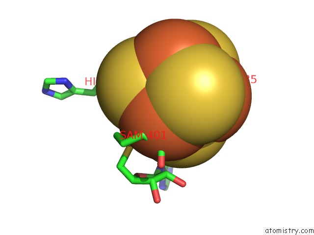
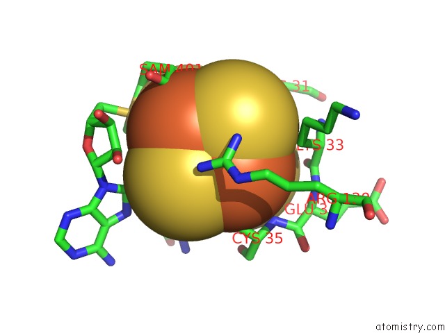
Iron binding site 3 out of 12 in 7n7i
Go back to
Iron binding site 3 out
of 12 in the X-Ray Crystal Structure of Viperin-Like Enzyme From Trichoderma Virens
Mono view
Stereo pair view
Mono view
Stereo pair view
A full contact list of Iron with other atoms in the Fe binding
site number 3 of X-Ray Crystal Structure of Viperin-Like Enzyme From Trichoderma Virens within 5.0Å range:
|
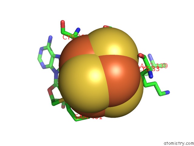
Iron binding site 4 out of 12 in 7n7i
Go back to
Iron binding site 4 out
of 12 in the X-Ray Crystal Structure of Viperin-Like Enzyme From Trichoderma Virens

Mono view
Stereo pair view
Mono view
Stereo pair view
A full contact list of Iron with other atoms in the Fe binding
site number 4 of X-Ray Crystal Structure of Viperin-Like Enzyme From Trichoderma Virens within 5.0Å range:
|
Iron binding site 5 out of 12 in 7n7i
Go back to
Iron binding site 5 out
of 12 in the X-Ray Crystal Structure of Viperin-Like Enzyme From Trichoderma Virens
Mono view
Stereo pair view
Mono view
Stereo pair view
A full contact list of Iron with other atoms in the Fe binding
site number 5 of X-Ray Crystal Structure of Viperin-Like Enzyme From Trichoderma Virens within 5.0Å range:
|
Iron binding site 6 out of 12 in 7n7i
Go back to
Iron binding site 6 out
of 12 in the X-Ray Crystal Structure of Viperin-Like Enzyme From Trichoderma Virens
Mono view
Stereo pair view
Mono view
Stereo pair view
A full contact list of Iron with other atoms in the Fe binding
site number 6 of X-Ray Crystal Structure of Viperin-Like Enzyme From Trichoderma Virens within 5.0Å range:
|
Iron binding site 7 out of 12 in 7n7i
Go back to
Iron binding site 7 out
of 12 in the X-Ray Crystal Structure of Viperin-Like Enzyme From Trichoderma Virens
Mono view
Stereo pair view
Mono view
Stereo pair view
A full contact list of Iron with other atoms in the Fe binding
site number 7 of X-Ray Crystal Structure of Viperin-Like Enzyme From Trichoderma Virens within 5.0Å range:
|
Iron binding site 8 out of 12 in 7n7i
Go back to
Iron binding site 8 out
of 12 in the X-Ray Crystal Structure of Viperin-Like Enzyme From Trichoderma Virens
Mono view
Stereo pair view
Mono view
Stereo pair view
A full contact list of Iron with other atoms in the Fe binding
site number 8 of X-Ray Crystal Structure of Viperin-Like Enzyme From Trichoderma Virens within 5.0Å range:
|
Iron binding site 9 out of 12 in 7n7i
Go back to
Iron binding site 9 out
of 12 in the X-Ray Crystal Structure of Viperin-Like Enzyme From Trichoderma Virens
Mono view
Stereo pair view
Mono view
Stereo pair view
A full contact list of Iron with other atoms in the Fe binding
site number 9 of X-Ray Crystal Structure of Viperin-Like Enzyme From Trichoderma Virens within 5.0Å range:
|
Iron binding site 10 out of 12 in 7n7i
Go back to
Iron binding site 10 out
of 12 in the X-Ray Crystal Structure of Viperin-Like Enzyme From Trichoderma Virens
Mono view
Stereo pair view
Mono view
Stereo pair view
A full contact list of Iron with other atoms in the Fe binding
site number 10 of X-Ray Crystal Structure of Viperin-Like Enzyme From Trichoderma Virens within 5.0Å range:
|
Reference:
J.C.Lachowicz,
A.G.Gizzi,
S.C.Almo,
T.L.Grove.
Structural Insight Into the Substrate Scope of Viperin and Viperin-Like Enzymes From Three Domains of Life To Be Published.
Page generated: Thu Aug 7 00:05:10 2025
Last articles
Mn in 9LJUMn in 9LJW
Mn in 9LJS
Mn in 9LJR
Mn in 9LJT
Mn in 9LJV
Mg in 9UA2
Mg in 9R96
Mg in 9VM1
Mg in 9P01