Iron »
PDB 7p7l-7pr2 »
7ppv »
Iron in PDB 7ppv: Structure of Dife-Sulerythrin at 2.70 Mgy Total Absorbed Dose
Protein crystallography data
The structure of Structure of Dife-Sulerythrin at 2.70 Mgy Total Absorbed Dose, PDB code: 7ppv
was solved by
F.Lennartz,
M.S.Weiss,
with X-Ray Crystallography technique. A brief refinement statistics is given in the table below:
| Resolution Low / High (Å) | 38.55 / 1.36 |
| Space group | P 63 |
| Cell size a, b, c (Å), α, β, γ (°) | 72.15, 72.15, 97.98, 90, 90, 120 |
| R / Rfree (%) | 18.1 / 19.9 |
Other elements in 7ppv:
The structure of Structure of Dife-Sulerythrin at 2.70 Mgy Total Absorbed Dose also contains other interesting chemical elements:
| Chlorine | (Cl) | 2 atoms |
Iron Binding Sites:
The binding sites of Iron atom in the Structure of Dife-Sulerythrin at 2.70 Mgy Total Absorbed Dose
(pdb code 7ppv). This binding sites where shown within
5.0 Angstroms radius around Iron atom.
In total 6 binding sites of Iron where determined in the Structure of Dife-Sulerythrin at 2.70 Mgy Total Absorbed Dose, PDB code: 7ppv:
Jump to Iron binding site number: 1; 2; 3; 4; 5; 6;
In total 6 binding sites of Iron where determined in the Structure of Dife-Sulerythrin at 2.70 Mgy Total Absorbed Dose, PDB code: 7ppv:
Jump to Iron binding site number: 1; 2; 3; 4; 5; 6;
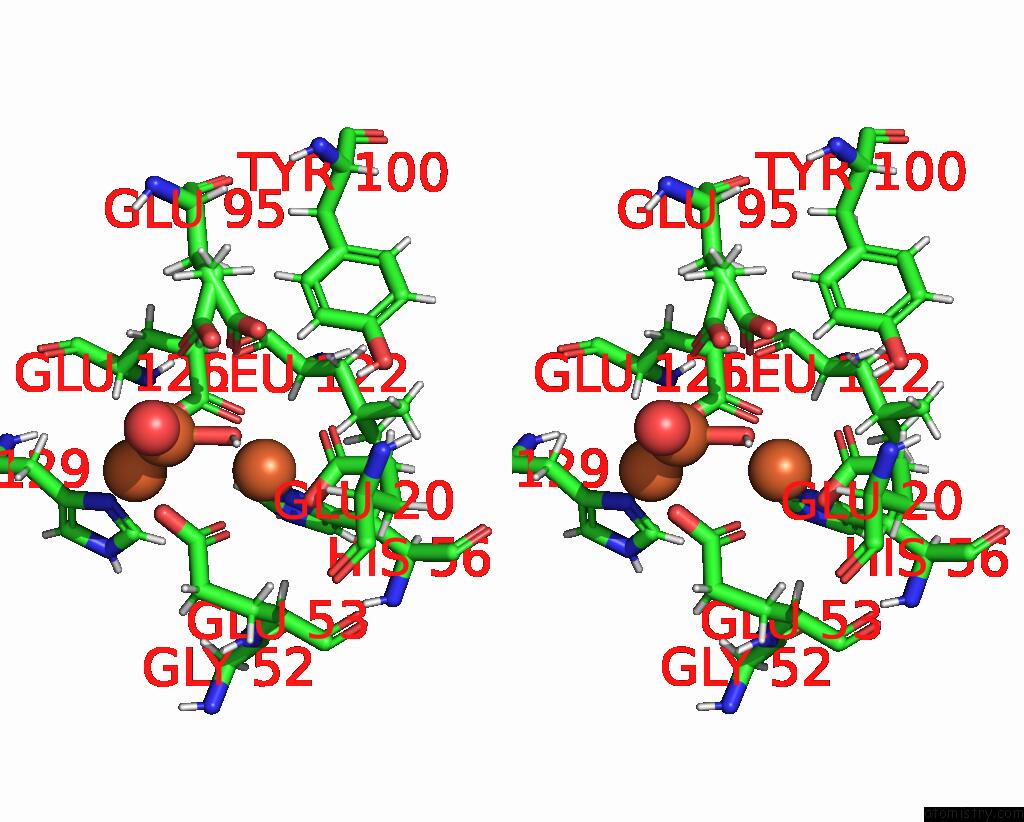
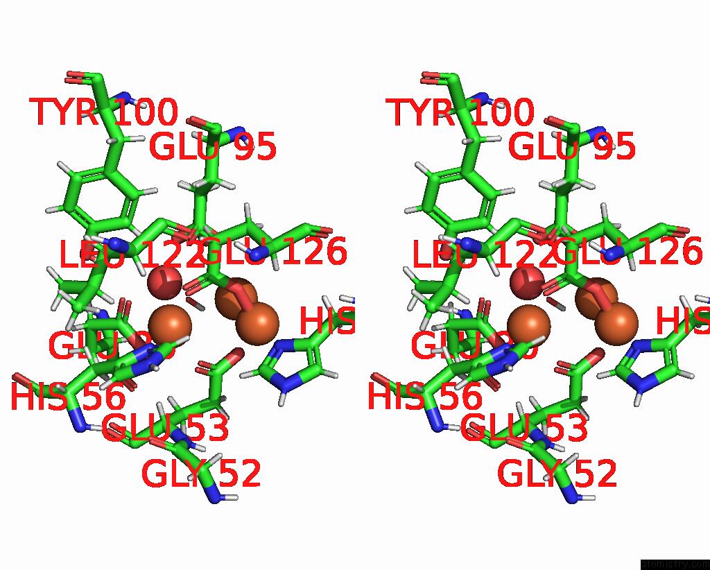
Iron binding site 1 out of 6 in 7ppv
Go back to
Iron binding site 1 out
of 6 in the Structure of Dife-Sulerythrin at 2.70 Mgy Total Absorbed Dose
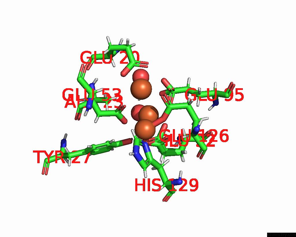
Mono view
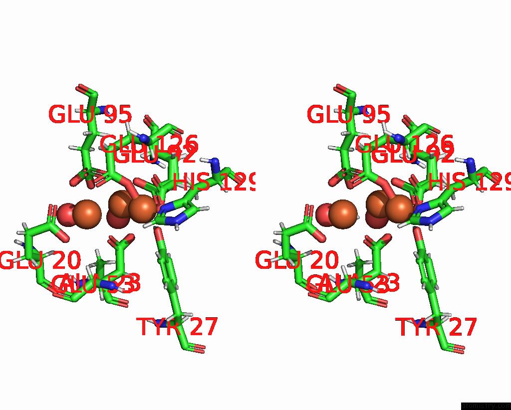
Stereo pair view
Mono view
Stereo pair view
A full contact list of Iron with other atoms in the Fe binding
site number 1 of Structure of Dife-Sulerythrin at 2.70 Mgy Total Absorbed Dose within 5.0Å range:
|
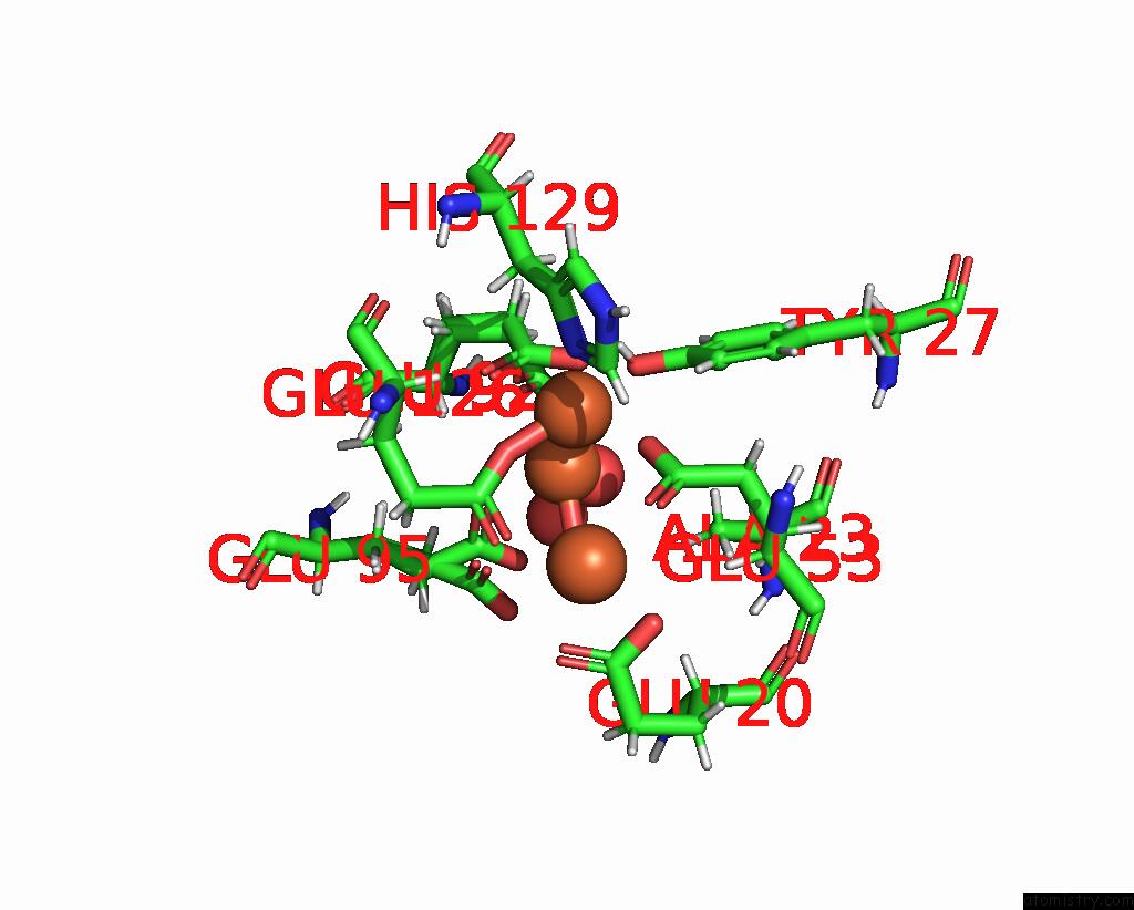
Iron binding site 2 out of 6 in 7ppv
Go back to
Iron binding site 2 out
of 6 in the Structure of Dife-Sulerythrin at 2.70 Mgy Total Absorbed Dose
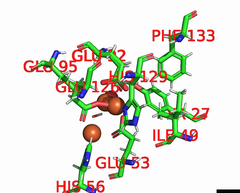
Mono view

Stereo pair view
Mono view
Stereo pair view
A full contact list of Iron with other atoms in the Fe binding
site number 2 of Structure of Dife-Sulerythrin at 2.70 Mgy Total Absorbed Dose within 5.0Å range:
|
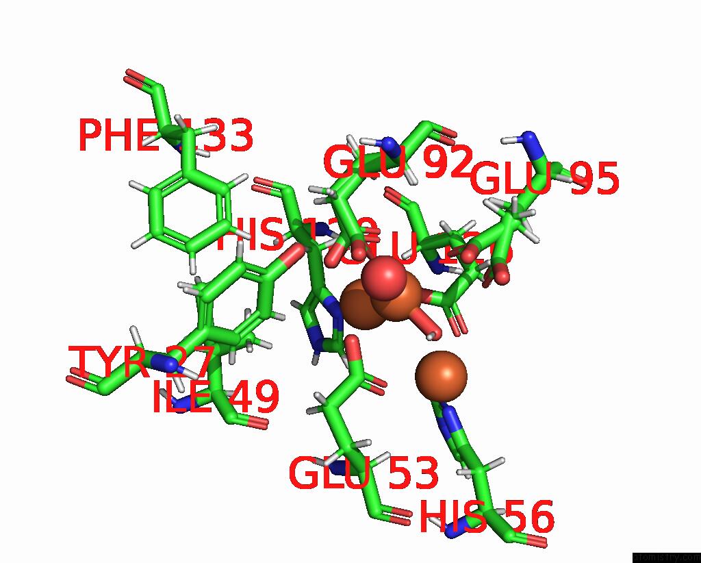
Iron binding site 3 out of 6 in 7ppv
Go back to
Iron binding site 3 out
of 6 in the Structure of Dife-Sulerythrin at 2.70 Mgy Total Absorbed Dose
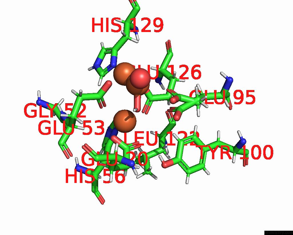
Mono view
Stereo pair view
Mono view
Stereo pair view
A full contact list of Iron with other atoms in the Fe binding
site number 3 of Structure of Dife-Sulerythrin at 2.70 Mgy Total Absorbed Dose within 5.0Å range:
|
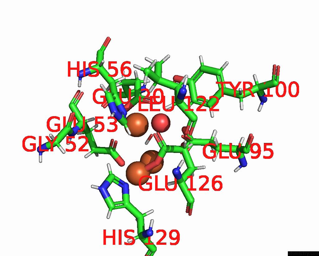
Iron binding site 4 out of 6 in 7ppv
Go back to
Iron binding site 4 out
of 6 in the Structure of Dife-Sulerythrin at 2.70 Mgy Total Absorbed Dose
Mono view

Stereo pair view
Mono view
Stereo pair view
A full contact list of Iron with other atoms in the Fe binding
site number 4 of Structure of Dife-Sulerythrin at 2.70 Mgy Total Absorbed Dose within 5.0Å range:
|
Iron binding site 5 out of 6 in 7ppv
Go back to
Iron binding site 5 out
of 6 in the Structure of Dife-Sulerythrin at 2.70 Mgy Total Absorbed Dose
Mono view

Stereo pair view
Mono view
Stereo pair view
A full contact list of Iron with other atoms in the Fe binding
site number 5 of Structure of Dife-Sulerythrin at 2.70 Mgy Total Absorbed Dose within 5.0Å range:
|
Iron binding site 6 out of 6 in 7ppv
Go back to
Iron binding site 6 out
of 6 in the Structure of Dife-Sulerythrin at 2.70 Mgy Total Absorbed Dose
Mono view
Stereo pair view
Mono view
Stereo pair view
A full contact list of Iron with other atoms in the Fe binding
site number 6 of Structure of Dife-Sulerythrin at 2.70 Mgy Total Absorbed Dose within 5.0Å range:
|
Reference:
F.Lennartz,
J.H.Jeoung,
S.Ruenger,
H.Dobbek,
M.S.Weiss.
Determining the Oxidation State of Elements By X-Ray Crystallography. Acta Crystallogr D Struct V. 78 238 2022BIOL.
ISSN: ISSN 2059-7983
PubMed: 35102889
DOI: 10.1107/S2059798321013048
Page generated: Thu Aug 7 02:55:26 2025
ISSN: ISSN 2059-7983
PubMed: 35102889
DOI: 10.1107/S2059798321013048
Last articles
Mg in 4DR7Mg in 4DR6
Mg in 4DR5
Mg in 4DUX
Mg in 4DUW
Mg in 4DUV
Mg in 4DUO
Mg in 4DUG
Mg in 4DTY
Mg in 4DTW