Iron »
PDB 7rl2-7sf6 »
7sbh »
Iron in PDB 7sbh: Crystal Structure of the Iron Superoxide Dismutase From Acinetobacter Sp. VER3
Enzymatic activity of Crystal Structure of the Iron Superoxide Dismutase From Acinetobacter Sp. VER3
All present enzymatic activity of Crystal Structure of the Iron Superoxide Dismutase From Acinetobacter Sp. VER3:
1.15.1.1;
1.15.1.1;
Protein crystallography data
The structure of Crystal Structure of the Iron Superoxide Dismutase From Acinetobacter Sp. VER3, PDB code: 7sbh
was solved by
B.A.Steimbruch,
D.Albanesi,
G.D.Repizo,
M.N.Lisa,
with X-Ray Crystallography technique. A brief refinement statistics is given in the table below:
| Resolution Low / High (Å) | 27.49 / 1.34 |
| Space group | C 2 2 21 |
| Cell size a, b, c (Å), α, β, γ (°) | 70.933, 87.016, 75.606, 90, 90, 90 |
| R / Rfree (%) | 14.4 / 16.2 |
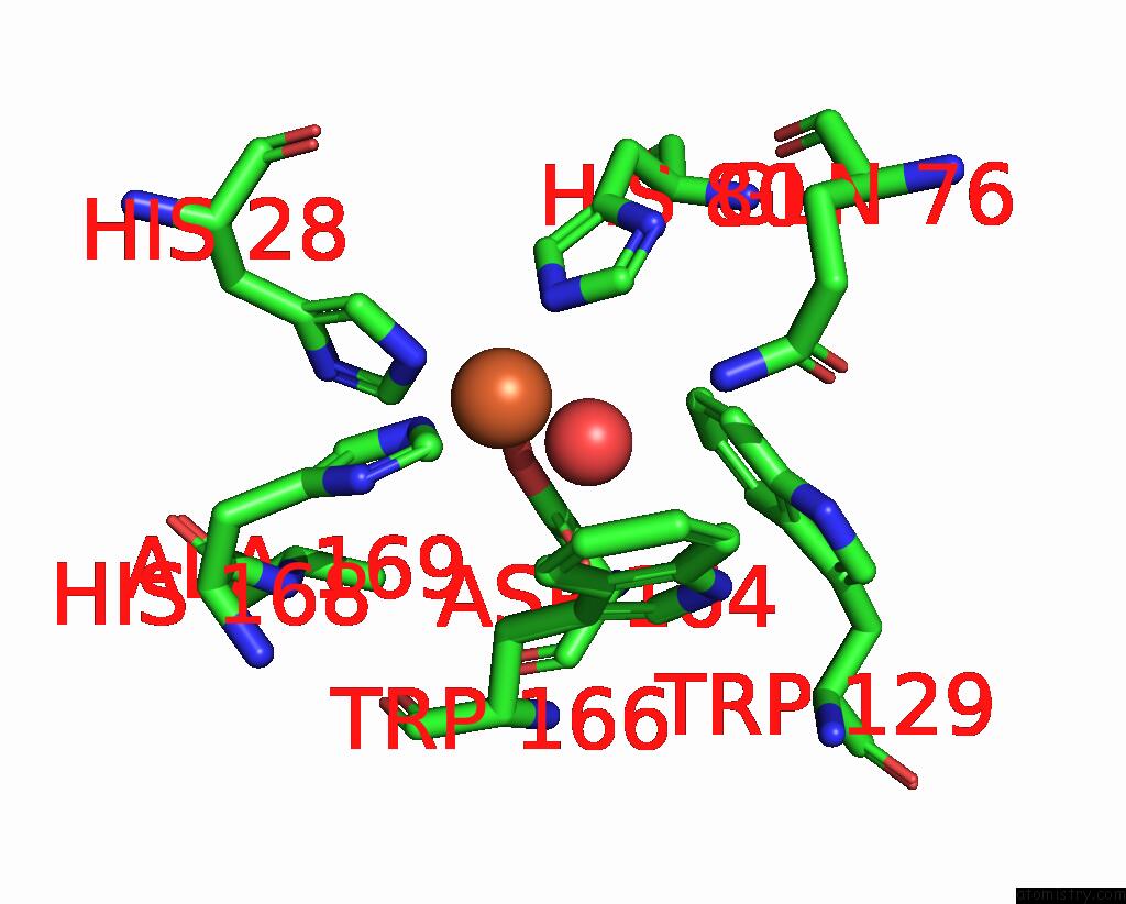
Iron Binding Sites:
The binding sites of Iron atom in the Crystal Structure of the Iron Superoxide Dismutase From Acinetobacter Sp. VER3
(pdb code 7sbh). This binding sites where shown within
5.0 Angstroms radius around Iron atom.
In total only one binding site of Iron was determined in the Crystal Structure of the Iron Superoxide Dismutase From Acinetobacter Sp. VER3, PDB code: 7sbh:
In total only one binding site of Iron was determined in the Crystal Structure of the Iron Superoxide Dismutase From Acinetobacter Sp. VER3, PDB code: 7sbh:
Iron binding site 1 out of 1 in 7sbh
Go back to
Iron binding site 1 out
of 1 in the Crystal Structure of the Iron Superoxide Dismutase From Acinetobacter Sp. VER3
Mono view

Stereo pair view
Mono view
Stereo pair view
A full contact list of Iron with other atoms in the Fe binding
site number 1 of Crystal Structure of the Iron Superoxide Dismutase From Acinetobacter Sp. VER3 within 5.0Å range:
|
Reference:
B.A.Steimbruch,
M.G.Sartorio,
N.Cortez,
D.Albanesi,
M.N.Lisa,
G.D.Repizo.
The Distinctive Roles Played By the Superoxide Dismutases of the Extremophile Acinetobacter Sp. VER3. Sci Rep V. 12 4321 2022.
ISSN: ESSN 2045-2322
PubMed: 35279679
DOI: 10.1038/S41598-022-08052-Z
Page generated: Thu Aug 7 05:30:33 2025
ISSN: ESSN 2045-2322
PubMed: 35279679
DOI: 10.1038/S41598-022-08052-Z
Last articles
Mn in 9LJUMn in 9LJW
Mn in 9LJS
Mn in 9LJR
Mn in 9LJT
Mn in 9LJV
Mg in 9UA2
Mg in 9R96
Mg in 9VM1
Mg in 9P01