Iron »
PDB 7tce-7tqh »
7thu »
Iron in PDB 7thu: Structure of Reduced Bovine Cytochrome C Oxidase at 1.93 Angstrom Resolution Obtained By Synchrotron X-Rays
Enzymatic activity of Structure of Reduced Bovine Cytochrome C Oxidase at 1.93 Angstrom Resolution Obtained By Synchrotron X-Rays
All present enzymatic activity of Structure of Reduced Bovine Cytochrome C Oxidase at 1.93 Angstrom Resolution Obtained By Synchrotron X-Rays:
1.9.3.1;
1.9.3.1;
Protein crystallography data
The structure of Structure of Reduced Bovine Cytochrome C Oxidase at 1.93 Angstrom Resolution Obtained By Synchrotron X-Rays, PDB code: 7thu
was solved by
I.Ishigami,
D.L.Rousseau,
S.-R.Yeh,
with X-Ray Crystallography technique. A brief refinement statistics is given in the table below:
| Resolution Low / High (Å) | 40.00 / 1.93 |
| Space group | P 21 21 21 |
| Cell size a, b, c (Å), α, β, γ (°) | 178.182, 182.282, 208.683, 90, 90, 90 |
| R / Rfree (%) | 18.4 / 21.3 |
Other elements in 7thu:
The structure of Structure of Reduced Bovine Cytochrome C Oxidase at 1.93 Angstrom Resolution Obtained By Synchrotron X-Rays also contains other interesting chemical elements:
| Zinc | (Zn) | 2 atoms |
| Copper | (Cu) | 6 atoms |
| Magnesium | (Mg) | 2 atoms |
| Sodium | (Na) | 2 atoms |
Iron Binding Sites:
The binding sites of Iron atom in the Structure of Reduced Bovine Cytochrome C Oxidase at 1.93 Angstrom Resolution Obtained By Synchrotron X-Rays
(pdb code 7thu). This binding sites where shown within
5.0 Angstroms radius around Iron atom.
In total 4 binding sites of Iron where determined in the Structure of Reduced Bovine Cytochrome C Oxidase at 1.93 Angstrom Resolution Obtained By Synchrotron X-Rays, PDB code: 7thu:
Jump to Iron binding site number: 1; 2; 3; 4;
In total 4 binding sites of Iron where determined in the Structure of Reduced Bovine Cytochrome C Oxidase at 1.93 Angstrom Resolution Obtained By Synchrotron X-Rays, PDB code: 7thu:
Jump to Iron binding site number: 1; 2; 3; 4;
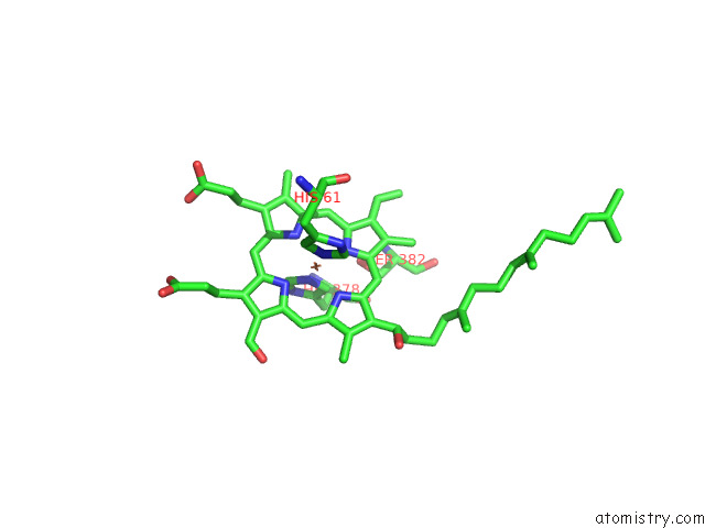
Iron binding site 1 out of 4 in 7thu
Go back to
Iron binding site 1 out
of 4 in the Structure of Reduced Bovine Cytochrome C Oxidase at 1.93 Angstrom Resolution Obtained By Synchrotron X-Rays

Mono view
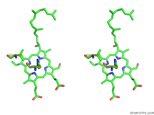
Stereo pair view
Mono view
Stereo pair view
|
|
A full contact list of Iron with other atoms in the Fe binding
site number 1 of Structure of Reduced Bovine Cytochrome C Oxidase at 1.93 Angstrom Resolution Obtained By Synchrotron X-Rays within 5.0Å range:
|
Iron binding site 2 out of 4 in 7thu
Go back to
Iron binding site 2 out
of 4 in the Structure of Reduced Bovine Cytochrome C Oxidase at 1.93 Angstrom Resolution Obtained By Synchrotron X-Rays

Mono view
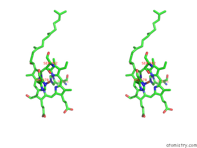
Stereo pair view
Mono view
Stereo pair view
|
|
A full contact list of Iron with other atoms in the Fe binding
site number 2 of Structure of Reduced Bovine Cytochrome C Oxidase at 1.93 Angstrom Resolution Obtained By Synchrotron X-Rays within 5.0Å range:
|
Iron binding site 3 out of 4 in 7thu
Go back to
Iron binding site 3 out
of 4 in the Structure of Reduced Bovine Cytochrome C Oxidase at 1.93 Angstrom Resolution Obtained By Synchrotron X-Rays
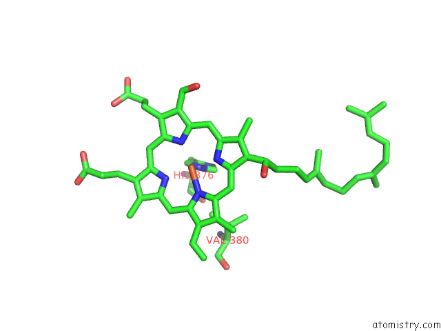
Mono view
Stereo pair view
Mono view
Stereo pair view
|
|
A full contact list of Iron with other atoms in the Fe binding
site number 3 of Structure of Reduced Bovine Cytochrome C Oxidase at 1.93 Angstrom Resolution Obtained By Synchrotron X-Rays within 5.0Å range:
|
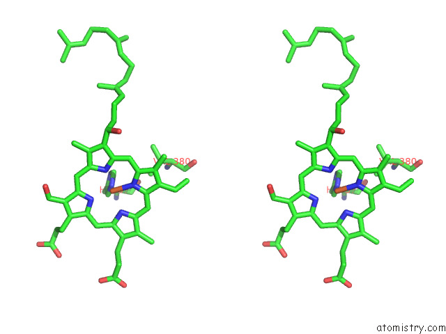
Iron binding site 4 out of 4 in 7thu
Go back to
Iron binding site 4 out
of 4 in the Structure of Reduced Bovine Cytochrome C Oxidase at 1.93 Angstrom Resolution Obtained By Synchrotron X-Rays
Mono view

Stereo pair view
Mono view
Stereo pair view
|
|
A full contact list of Iron with other atoms in the Fe binding
site number 4 of Structure of Reduced Bovine Cytochrome C Oxidase at 1.93 Angstrom Resolution Obtained By Synchrotron X-Rays within 5.0Å range:
|
Reference:
I.Ishigami,
S.Russi,
A.Cohen,
S.R.Yeh,
D.L.Rousseau.
Temperature-Dependent Structural Transition Following X-Ray-Induced Metal Center Reduction in Oxidized Cytochrome C Oxidase. J.Biol.Chem. V. 298 01799 2022.
ISSN: ESSN 1083-351X
PubMed: 35257742
DOI: 10.1016/J.JBC.2022.101799
Page generated: Thu Aug 7 05:56:00 2025
ISSN: ESSN 1083-351X
PubMed: 35257742
DOI: 10.1016/J.JBC.2022.101799
Last articles
Ni in 446DNi in 3ZG1
Ni in 3ZR9
Ni in 3ZUC
Ni in 3ZU8
Ni in 3ZQW
Ni in 3ZF5
Ni in 3ZF6
Ni in 3ZF4
Ni in 3ZFS