Iron »
PDB 7txe-7us8 »
7ud7 »
Iron in PDB 7ud7: Crystal Structure of Deoxygenated Hemoglobin in Complex with 5HMF-No at 1.8 Angstrom
Protein crystallography data
The structure of Crystal Structure of Deoxygenated Hemoglobin in Complex with 5HMF-No at 1.8 Angstrom, PDB code: 7ud7
was solved by
A.K.Donkor,
F.N.Musayev,
M.K.Safo,
with X-Ray Crystallography technique. A brief refinement statistics is given in the table below:
| Resolution Low / High (Å) | 26.36 / 1.80 |
| Space group | P 1 21 1 |
| Cell size a, b, c (Å), α, β, γ (°) | 62.344, 82.031, 53.454, 90, 99.49, 90 |
| R / Rfree (%) | 15.1 / 19.1 |
Iron Binding Sites:
The binding sites of Iron atom in the Crystal Structure of Deoxygenated Hemoglobin in Complex with 5HMF-No at 1.8 Angstrom
(pdb code 7ud7). This binding sites where shown within
5.0 Angstroms radius around Iron atom.
In total 4 binding sites of Iron where determined in the Crystal Structure of Deoxygenated Hemoglobin in Complex with 5HMF-No at 1.8 Angstrom, PDB code: 7ud7:
Jump to Iron binding site number: 1; 2; 3; 4;
In total 4 binding sites of Iron where determined in the Crystal Structure of Deoxygenated Hemoglobin in Complex with 5HMF-No at 1.8 Angstrom, PDB code: 7ud7:
Jump to Iron binding site number: 1; 2; 3; 4;
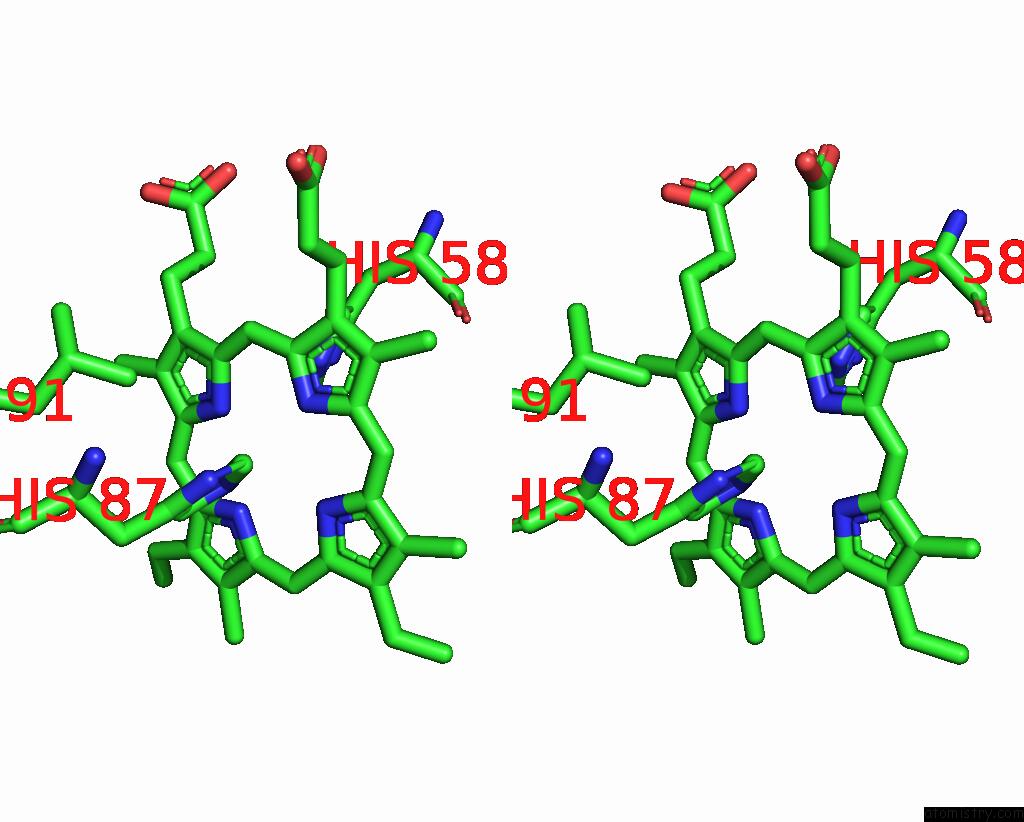
Iron binding site 1 out of 4 in 7ud7
Go back to
Iron binding site 1 out
of 4 in the Crystal Structure of Deoxygenated Hemoglobin in Complex with 5HMF-No at 1.8 Angstrom
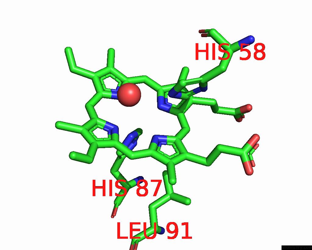
Mono view
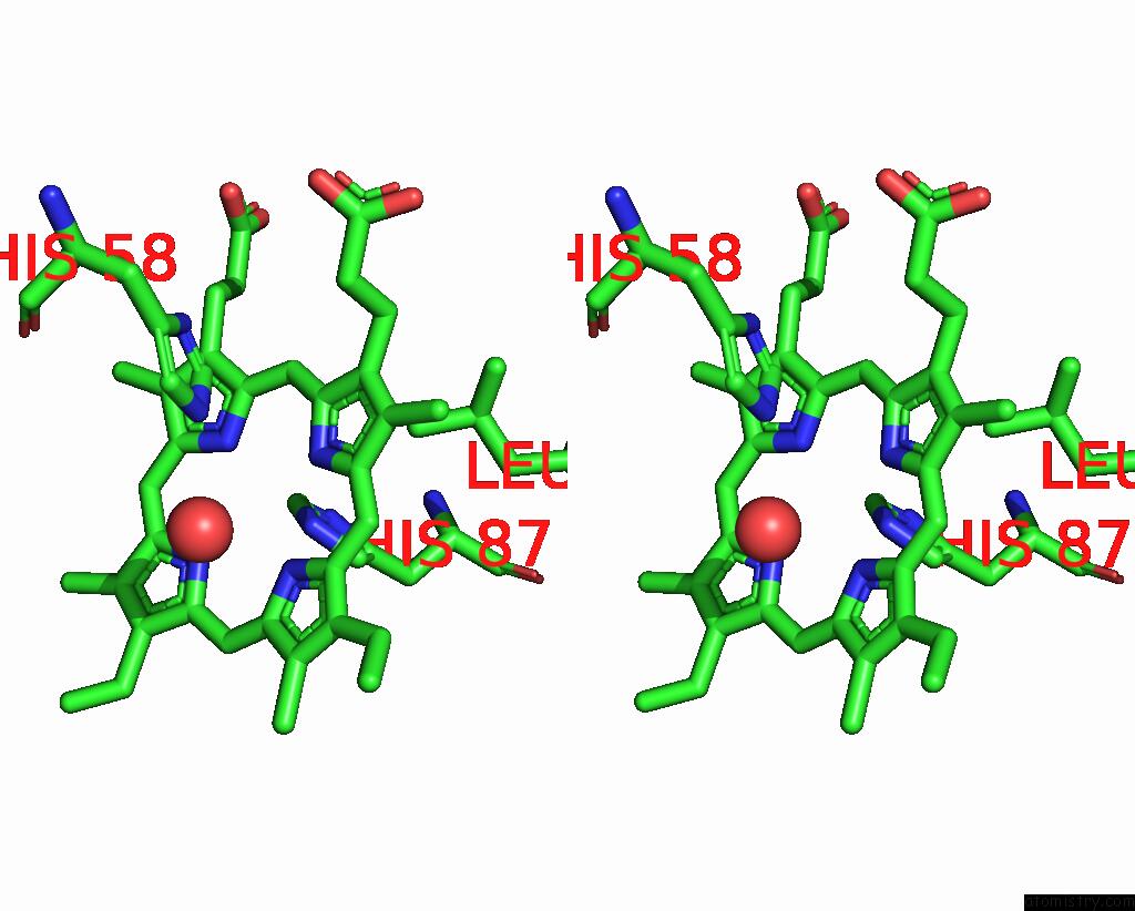
Stereo pair view
Mono view
Stereo pair view
A full contact list of Iron with other atoms in the Fe binding
site number 1 of Crystal Structure of Deoxygenated Hemoglobin in Complex with 5HMF-No at 1.8 Angstrom within 5.0Å range:
|
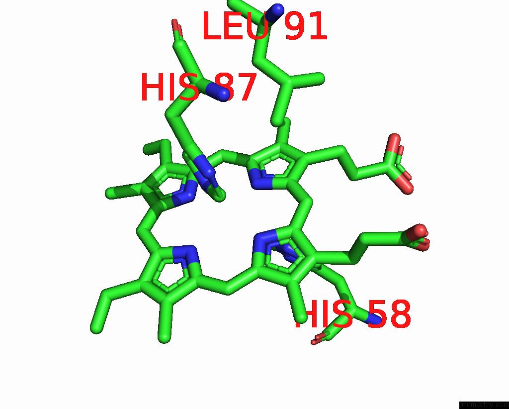
Iron binding site 2 out of 4 in 7ud7
Go back to
Iron binding site 2 out
of 4 in the Crystal Structure of Deoxygenated Hemoglobin in Complex with 5HMF-No at 1.8 Angstrom

Mono view
Stereo pair view
Mono view
Stereo pair view
A full contact list of Iron with other atoms in the Fe binding
site number 2 of Crystal Structure of Deoxygenated Hemoglobin in Complex with 5HMF-No at 1.8 Angstrom within 5.0Å range:
|
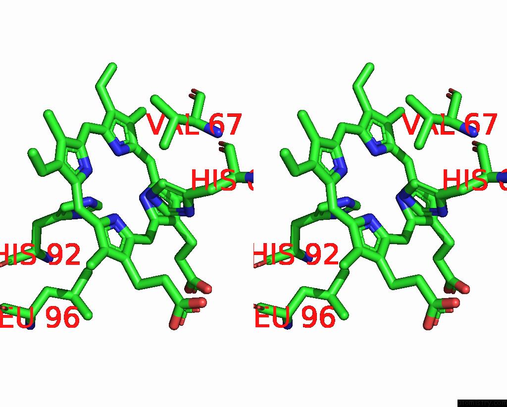
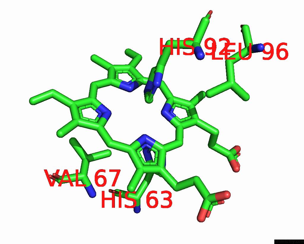
Iron binding site 3 out of 4 in 7ud7
Go back to
Iron binding site 3 out
of 4 in the Crystal Structure of Deoxygenated Hemoglobin in Complex with 5HMF-No at 1.8 Angstrom
Mono view
Stereo pair view
Mono view
Stereo pair view
A full contact list of Iron with other atoms in the Fe binding
site number 3 of Crystal Structure of Deoxygenated Hemoglobin in Complex with 5HMF-No at 1.8 Angstrom within 5.0Å range:
|
Iron binding site 4 out of 4 in 7ud7
Go back to
Iron binding site 4 out
of 4 in the Crystal Structure of Deoxygenated Hemoglobin in Complex with 5HMF-No at 1.8 Angstrom
Mono view

Stereo pair view
Mono view
Stereo pair view
A full contact list of Iron with other atoms in the Fe binding
site number 4 of Crystal Structure of Deoxygenated Hemoglobin in Complex with 5HMF-No at 1.8 Angstrom within 5.0Å range:
|
Reference:
R.T.Alhashimi,
M.S.Ghatge,
A.K.Donkor,
T.M.Deshpande,
N.Anabaraonye,
D.Alramadhani,
R.Danso-Danquah,
B.Huang,
Y.Zhang,
F.N.Musayev,
O.Abdulmalik,
M.K.Safo.
Design, Synthesis, and Antisickling Investigation of A Nitric Oxide-Releasing Prodrug of 5HMF For the Treatment of Sickle Cell Disease. Biomolecules V. 12 2022.
ISSN: ESSN 2218-273X
PubMed: 35625623
DOI: 10.3390/BIOM12050696
Page generated: Thu Aug 7 06:19:31 2025
ISSN: ESSN 2218-273X
PubMed: 35625623
DOI: 10.3390/BIOM12050696
Last articles
Sr in 3BNQSr in 2QJY
Sr in 2X53
Sr in 2SPT
Sr in 2XRM
Sr in 2WOH
Sr in 2RIO
Sr in 2PN4
Sr in 2QJK
Sr in 2QJP