Iron »
PDB 7wdh-7xgy »
7wdi »
Iron in PDB 7wdi: Crystal Structure of the P450 BM3 Heme Domain Mutant F87K in Complex with N-Imidazolyl-Hexanoyl-L-Phenylalanine and Hydroxylamine
Enzymatic activity of Crystal Structure of the P450 BM3 Heme Domain Mutant F87K in Complex with N-Imidazolyl-Hexanoyl-L-Phenylalanine and Hydroxylamine
All present enzymatic activity of Crystal Structure of the P450 BM3 Heme Domain Mutant F87K in Complex with N-Imidazolyl-Hexanoyl-L-Phenylalanine and Hydroxylamine:
1.14.14.1; 1.6.2.4;
1.14.14.1; 1.6.2.4;
Protein crystallography data
The structure of Crystal Structure of the P450 BM3 Heme Domain Mutant F87K in Complex with N-Imidazolyl-Hexanoyl-L-Phenylalanine and Hydroxylamine, PDB code: 7wdi
was solved by
Y.Jiang,
S.Dong,
Y.Feng,
Z.Cong,
with X-Ray Crystallography technique. A brief refinement statistics is given in the table below:
| Resolution Low / High (Å) | 29.25 / 2.10 |
| Space group | P 1 21 1 |
| Cell size a, b, c (Å), α, β, γ (°) | 58.413, 149.017, 65.281, 90, 100.35, 90 |
| R / Rfree (%) | 18.4 / 21.9 |
Iron Binding Sites:
The binding sites of Iron atom in the Crystal Structure of the P450 BM3 Heme Domain Mutant F87K in Complex with N-Imidazolyl-Hexanoyl-L-Phenylalanine and Hydroxylamine
(pdb code 7wdi). This binding sites where shown within
5.0 Angstroms radius around Iron atom.
In total 2 binding sites of Iron where determined in the Crystal Structure of the P450 BM3 Heme Domain Mutant F87K in Complex with N-Imidazolyl-Hexanoyl-L-Phenylalanine and Hydroxylamine, PDB code: 7wdi:
Jump to Iron binding site number: 1; 2;
In total 2 binding sites of Iron where determined in the Crystal Structure of the P450 BM3 Heme Domain Mutant F87K in Complex with N-Imidazolyl-Hexanoyl-L-Phenylalanine and Hydroxylamine, PDB code: 7wdi:
Jump to Iron binding site number: 1; 2;
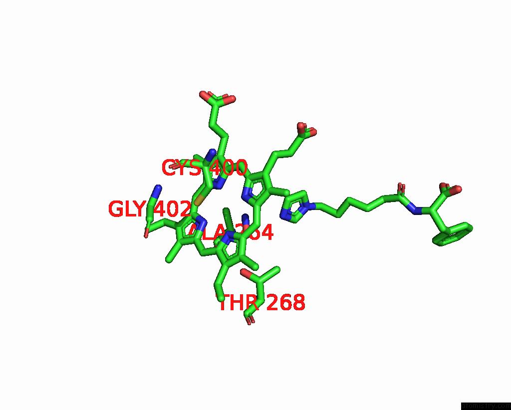
Iron binding site 1 out of 2 in 7wdi
Go back to
Iron binding site 1 out
of 2 in the Crystal Structure of the P450 BM3 Heme Domain Mutant F87K in Complex with N-Imidazolyl-Hexanoyl-L-Phenylalanine and Hydroxylamine

Mono view
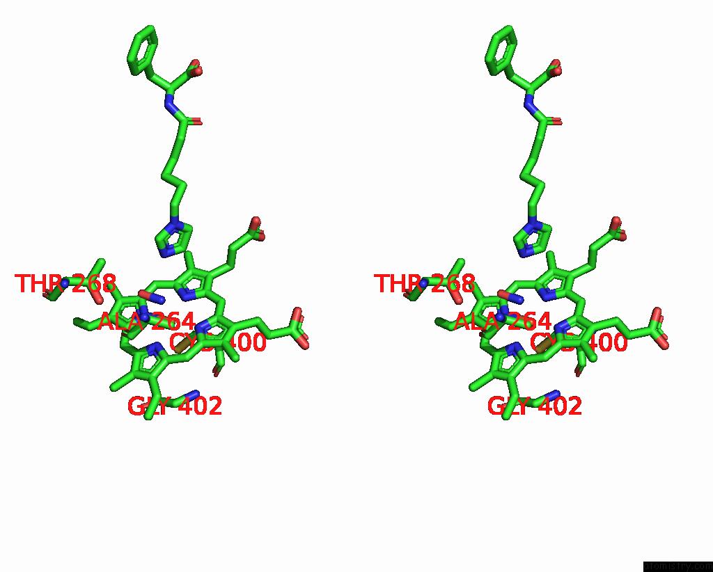
Stereo pair view
Mono view
Stereo pair view
A full contact list of Iron with other atoms in the Fe binding
site number 1 of Crystal Structure of the P450 BM3 Heme Domain Mutant F87K in Complex with N-Imidazolyl-Hexanoyl-L-Phenylalanine and Hydroxylamine within 5.0Å range:
|
Iron binding site 2 out of 2 in 7wdi
Go back to
Iron binding site 2 out
of 2 in the Crystal Structure of the P450 BM3 Heme Domain Mutant F87K in Complex with N-Imidazolyl-Hexanoyl-L-Phenylalanine and Hydroxylamine
Mono view
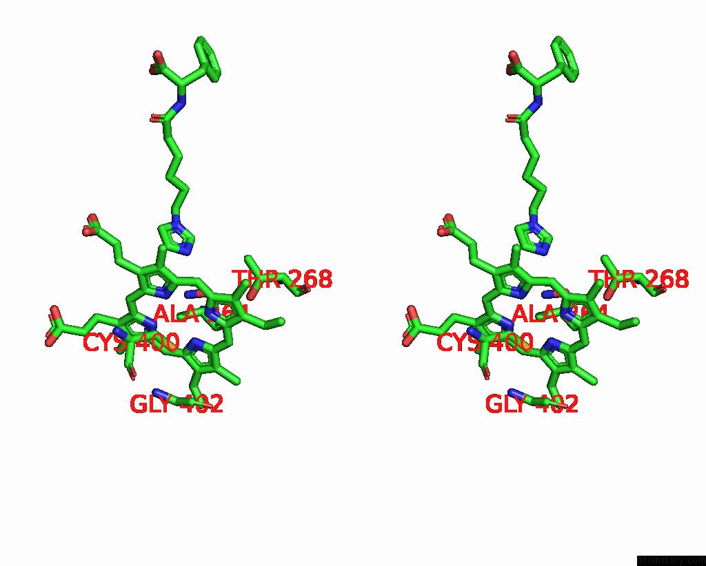
Stereo pair view
Mono view
Stereo pair view
A full contact list of Iron with other atoms in the Fe binding
site number 2 of Crystal Structure of the P450 BM3 Heme Domain Mutant F87K in Complex with N-Imidazolyl-Hexanoyl-L-Phenylalanine and Hydroxylamine within 5.0Å range:
|
Reference:
Y.Jiang,
Z.Cong.
Crystal Structure of the P450 BM3 Heme Domain Mutant F87A in Complex with N-Imidazolyl-Hexanoyl-L-Phenylalanine and Hydroxylamine To Be Published.
Page generated: Thu Aug 7 09:51:43 2025
Last articles
Xe in 1UX9Xe in 1VGI
Xe in 1URY
Xe in 1VAU
Xe in 1UVX
Xe in 1S56
Xe in 1UVY
Xe in 1UOC
Xe in 1UO6
Xe in 1U0X