Iron »
PDB 7wdh-7xgy »
7wzm »
Iron in PDB 7wzm: Crystal Structure of Cytochrome P450 184A1 From Streptomyces Avermitilis in Complex with Oleic Acid
Protein crystallography data
The structure of Crystal Structure of Cytochrome P450 184A1 From Streptomyces Avermitilis in Complex with Oleic Acid, PDB code: 7wzm
was solved by
V.C.Kim,
D.G.Kim,
S.G.Lee,
G.H.Lee,
S.A.Lee,
L.W.Kang,
with X-Ray Crystallography technique. A brief refinement statistics is given in the table below:
| Resolution Low / High (Å) | 45.52 / 1.68 |
| Space group | P 21 21 21 |
| Cell size a, b, c (Å), α, β, γ (°) | 54.687, 62.652, 132.228, 90, 90, 90 |
| R / Rfree (%) | 18.8 / 22.1 |
Iron Binding Sites:
The binding sites of Iron atom in the Crystal Structure of Cytochrome P450 184A1 From Streptomyces Avermitilis in Complex with Oleic Acid
(pdb code 7wzm). This binding sites where shown within
5.0 Angstroms radius around Iron atom.
In total only one binding site of Iron was determined in the Crystal Structure of Cytochrome P450 184A1 From Streptomyces Avermitilis in Complex with Oleic Acid, PDB code: 7wzm:
In total only one binding site of Iron was determined in the Crystal Structure of Cytochrome P450 184A1 From Streptomyces Avermitilis in Complex with Oleic Acid, PDB code: 7wzm:
Iron binding site 1 out of 1 in 7wzm
Go back to
Iron binding site 1 out
of 1 in the Crystal Structure of Cytochrome P450 184A1 From Streptomyces Avermitilis in Complex with Oleic Acid

Mono view
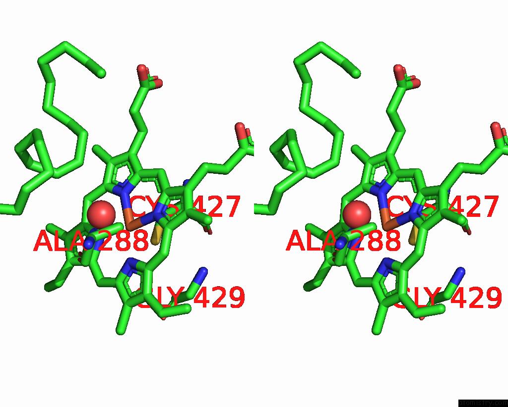
Stereo pair view
Mono view
Stereo pair view
A full contact list of Iron with other atoms in the Fe binding
site number 1 of Crystal Structure of Cytochrome P450 184A1 From Streptomyces Avermitilis in Complex with Oleic Acid within 5.0Å range:
|
Reference:
V.C.Kim,
D.G.Kim,
S.G.Lee,
G.H.Lee,
S.A.Lee,
L.W.Kang.
Crystal Structure of Cytochrome P450 184A1 From Streptomyces Avermitilis in Complex with Oleic Acid To Be Published.
Page generated: Thu Aug 7 09:56:29 2025
Last articles
Xe in 1FZIXe in 1I4W
Xe in 1GKZ
Xe in 1FO6
Xe in 1C6N
Xe in 1C6K
Xe in 1C6T
Xe in 1C6H
Xe in 1C6E
Xe in 1C68