Iron »
PDB 7xk3-7y9k »
7y3u »
Iron in PDB 7y3u: Crystal Structure of the Complex of Lactoperoxidase with Nitric Oxide at 2.50A Resolution
Enzymatic activity of Crystal Structure of the Complex of Lactoperoxidase with Nitric Oxide at 2.50A Resolution
All present enzymatic activity of Crystal Structure of the Complex of Lactoperoxidase with Nitric Oxide at 2.50A Resolution:
1.11.1.7;
1.11.1.7;
Protein crystallography data
The structure of Crystal Structure of the Complex of Lactoperoxidase with Nitric Oxide at 2.50A Resolution, PDB code: 7y3u
was solved by
P.K.Singh,
V.Viswanathan,
N.Ahmad,
C.Rani,
P.Sharma,
S.Sharma,
T.P.Singh,
with X-Ray Crystallography technique. A brief refinement statistics is given in the table below:
| Resolution Low / High (Å) | 48.86 / 2.50 |
| Space group | P 1 21 1 |
| Cell size a, b, c (Å), α, β, γ (°) | 81.731, 93.173, 82.319, 90, 91.2, 90 |
| R / Rfree (%) | 21.9 / 27.7 |
Other elements in 7y3u:
The structure of Crystal Structure of the Complex of Lactoperoxidase with Nitric Oxide at 2.50A Resolution also contains other interesting chemical elements:
| Calcium | (Ca) | 2 atoms |
| Chlorine | (Cl) | 1 atom |
Iron Binding Sites:
The binding sites of Iron atom in the Crystal Structure of the Complex of Lactoperoxidase with Nitric Oxide at 2.50A Resolution
(pdb code 7y3u). This binding sites where shown within
5.0 Angstroms radius around Iron atom.
In total 2 binding sites of Iron where determined in the Crystal Structure of the Complex of Lactoperoxidase with Nitric Oxide at 2.50A Resolution, PDB code: 7y3u:
Jump to Iron binding site number: 1; 2;
In total 2 binding sites of Iron where determined in the Crystal Structure of the Complex of Lactoperoxidase with Nitric Oxide at 2.50A Resolution, PDB code: 7y3u:
Jump to Iron binding site number: 1; 2;
Iron binding site 1 out of 2 in 7y3u
Go back to
Iron binding site 1 out
of 2 in the Crystal Structure of the Complex of Lactoperoxidase with Nitric Oxide at 2.50A Resolution
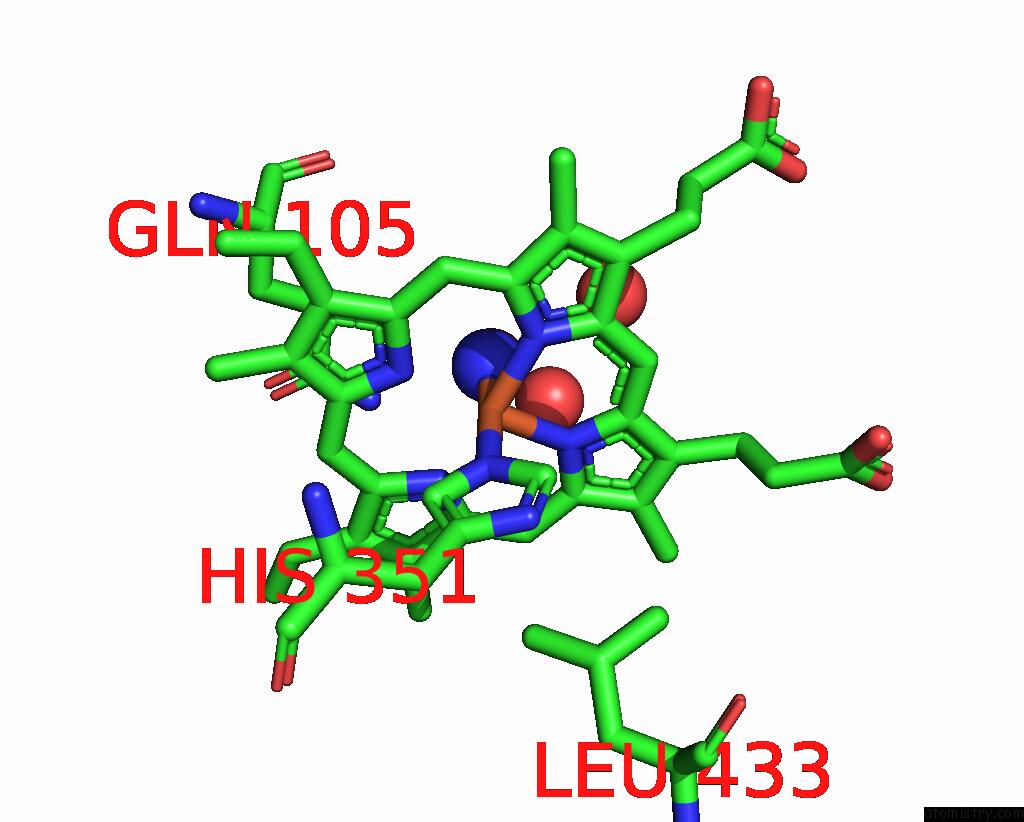
Mono view
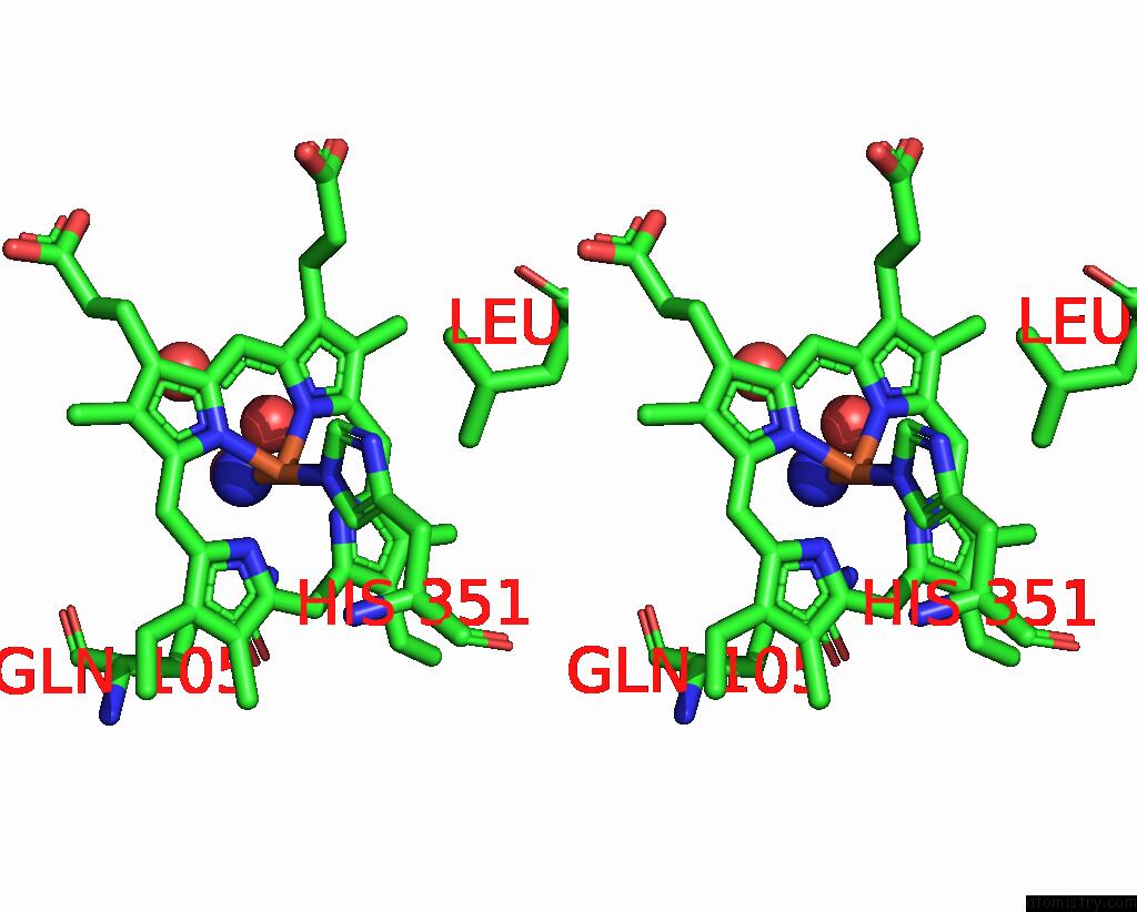
Stereo pair view
Mono view
Stereo pair view
A full contact list of Iron with other atoms in the Fe binding
site number 1 of Crystal Structure of the Complex of Lactoperoxidase with Nitric Oxide at 2.50A Resolution within 5.0Å range:
|
Iron binding site 2 out of 2 in 7y3u
Go back to
Iron binding site 2 out
of 2 in the Crystal Structure of the Complex of Lactoperoxidase with Nitric Oxide at 2.50A Resolution
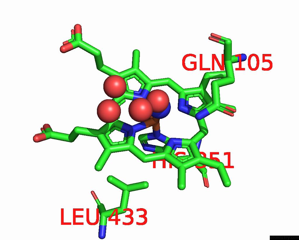
Mono view

Stereo pair view
Mono view
Stereo pair view
A full contact list of Iron with other atoms in the Fe binding
site number 2 of Crystal Structure of the Complex of Lactoperoxidase with Nitric Oxide at 2.50A Resolution within 5.0Å range:
|
Reference:
P.K.Singh,
V.Viswanathan,
N.Ahmad.
Crystal Structure of the Complex of Lactoperoxidase with Nitric Oxide at 2.50A Resolution To Be Published.
Page generated: Thu Aug 7 10:13:17 2025
Last articles
Na in 2QB4Na in 2QD6
Na in 2QCI
Na in 2QA0
Na in 2Q95
Na in 2Q96
Na in 2Q94
Na in 2Q93
Na in 2Q8X
Na in 2Q72