Iron »
PDB 7xk3-7y9k »
7y3y »
Iron in PDB 7y3y: Crystal Structure of BTG13 Mutant (T299V)
Protein crystallography data
The structure of Crystal Structure of BTG13 Mutant (T299V), PDB code: 7y3y
was solved by
X.D.Hou,
K.Fu,
Y.J.Rao,
with X-Ray Crystallography technique. A brief refinement statistics is given in the table below:
| Resolution Low / High (Å) | 78.99 / 1.92 |
| Space group | P 21 21 21 |
| Cell size a, b, c (Å), α, β, γ (°) | 79.31, 99.558, 129.781, 90, 90, 90 |
| R / Rfree (%) | 17 / 20.1 |
Iron Binding Sites:
The binding sites of Iron atom in the Crystal Structure of BTG13 Mutant (T299V)
(pdb code 7y3y). This binding sites where shown within
5.0 Angstroms radius around Iron atom.
In total 2 binding sites of Iron where determined in the Crystal Structure of BTG13 Mutant (T299V), PDB code: 7y3y:
Jump to Iron binding site number: 1; 2;
In total 2 binding sites of Iron where determined in the Crystal Structure of BTG13 Mutant (T299V), PDB code: 7y3y:
Jump to Iron binding site number: 1; 2;
Iron binding site 1 out of 2 in 7y3y
Go back to
Iron binding site 1 out
of 2 in the Crystal Structure of BTG13 Mutant (T299V)
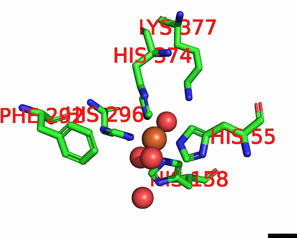
Mono view
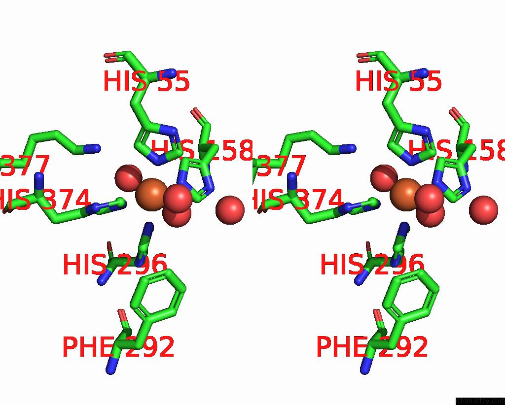
Stereo pair view
Mono view
Stereo pair view
A full contact list of Iron with other atoms in the Fe binding
site number 1 of Crystal Structure of BTG13 Mutant (T299V) within 5.0Å range:
|
Iron binding site 2 out of 2 in 7y3y
Go back to
Iron binding site 2 out
of 2 in the Crystal Structure of BTG13 Mutant (T299V)
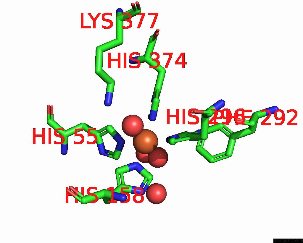
Mono view

Stereo pair view
Mono view
Stereo pair view
A full contact list of Iron with other atoms in the Fe binding
site number 2 of Crystal Structure of BTG13 Mutant (T299V) within 5.0Å range:
|
Reference:
X.Hou,
H.Xu,
Z.Deng,
Y.Yan,
Z.Yuan,
X.Liu,
Z.Su,
S.Yang,
Y.Zhang,
Y.Rao.
Discovery of the Biosynthetic Pathway of Beticolin 1 Reveals A Novel Non-Heme Iron-Dependent Oxygenase For Anthraquinone Ring Cleavage. Angew.Chem.Int.Ed.Engl. V. 61 08772 2022.
ISSN: ESSN 1521-3773
PubMed: 35862137
DOI: 10.1002/ANIE.202208772
Page generated: Thu Aug 7 10:14:03 2025
ISSN: ESSN 1521-3773
PubMed: 35862137
DOI: 10.1002/ANIE.202208772
Last articles
Mg in 1VPAMg in 1VPE
Mg in 1VOM
Mg in 1VMA
Mg in 1VMK
Mg in 1VM9
Mg in 1VCR
Mg in 1VLB
Mg in 1VKP
Mg in 1VL8