Iron »
PDB 7y9l-7yzt »
7y9l »
Iron in PDB 7y9l: Crystal Structure of P450 BM3-2F From Bacillus Megaterium in Complex with 2-Hydroxy-5-Nitrobenzonitrile
Enzymatic activity of Crystal Structure of P450 BM3-2F From Bacillus Megaterium in Complex with 2-Hydroxy-5-Nitrobenzonitrile
All present enzymatic activity of Crystal Structure of P450 BM3-2F From Bacillus Megaterium in Complex with 2-Hydroxy-5-Nitrobenzonitrile:
1.14.14.1; 1.6.2.4;
1.14.14.1; 1.6.2.4;
Protein crystallography data
The structure of Crystal Structure of P450 BM3-2F From Bacillus Megaterium in Complex with 2-Hydroxy-5-Nitrobenzonitrile, PDB code: 7y9l
was solved by
Q.Wang,
L.L.Zhang,
W.D.Liu,
J.-W.Huang,
Y.Yang,
C.-C.Chen,
R.-T.Guo,
with X-Ray Crystallography technique. A brief refinement statistics is given in the table below:
| Resolution Low / High (Å) | 24.78 / 1.76 |
| Space group | P 1 21 1 |
| Cell size a, b, c (Å), α, β, γ (°) | 59.014, 146.956, 63.271, 90, 98.07, 90 |
| R / Rfree (%) | 17.3 / 20.3 |
Other elements in 7y9l:
The structure of Crystal Structure of P450 BM3-2F From Bacillus Megaterium in Complex with 2-Hydroxy-5-Nitrobenzonitrile also contains other interesting chemical elements:
| Nickel | (Ni) | 2 atoms |
Iron Binding Sites:
The binding sites of Iron atom in the Crystal Structure of P450 BM3-2F From Bacillus Megaterium in Complex with 2-Hydroxy-5-Nitrobenzonitrile
(pdb code 7y9l). This binding sites where shown within
5.0 Angstroms radius around Iron atom.
In total 2 binding sites of Iron where determined in the Crystal Structure of P450 BM3-2F From Bacillus Megaterium in Complex with 2-Hydroxy-5-Nitrobenzonitrile, PDB code: 7y9l:
Jump to Iron binding site number: 1; 2;
In total 2 binding sites of Iron where determined in the Crystal Structure of P450 BM3-2F From Bacillus Megaterium in Complex with 2-Hydroxy-5-Nitrobenzonitrile, PDB code: 7y9l:
Jump to Iron binding site number: 1; 2;
Iron binding site 1 out of 2 in 7y9l
Go back to
Iron binding site 1 out
of 2 in the Crystal Structure of P450 BM3-2F From Bacillus Megaterium in Complex with 2-Hydroxy-5-Nitrobenzonitrile
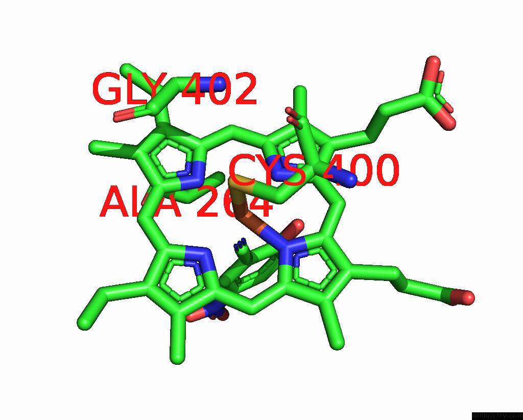
Mono view
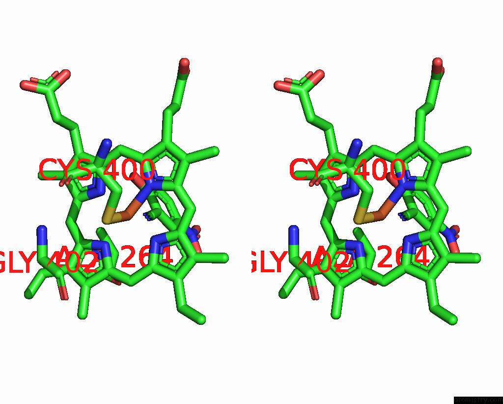
Stereo pair view
Mono view
Stereo pair view
A full contact list of Iron with other atoms in the Fe binding
site number 1 of Crystal Structure of P450 BM3-2F From Bacillus Megaterium in Complex with 2-Hydroxy-5-Nitrobenzonitrile within 5.0Å range:
|
Iron binding site 2 out of 2 in 7y9l
Go back to
Iron binding site 2 out
of 2 in the Crystal Structure of P450 BM3-2F From Bacillus Megaterium in Complex with 2-Hydroxy-5-Nitrobenzonitrile
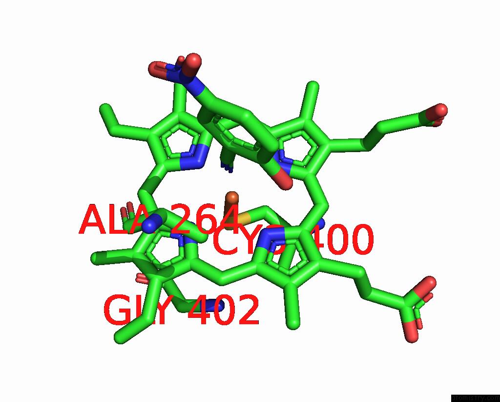
Mono view
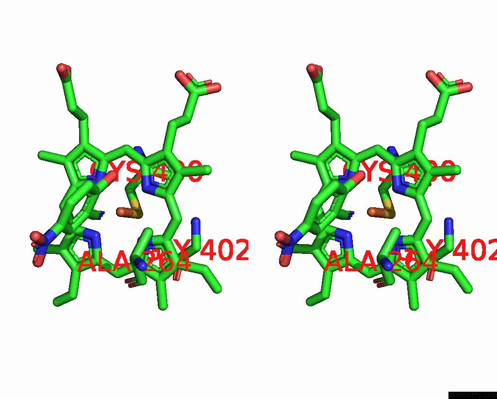
Stereo pair view
Mono view
Stereo pair view
A full contact list of Iron with other atoms in the Fe binding
site number 2 of Crystal Structure of P450 BM3-2F From Bacillus Megaterium in Complex with 2-Hydroxy-5-Nitrobenzonitrile within 5.0Å range:
|
Reference:
Q.Wang,
L.L.Zhang,
W.D.Liu,
J.-W.Huang,
Y.Yang,
C.-C.Chen,
R.-T.Guo.
Crystal Structure of P450 BM3-2F From Bacillus Megaterium in Complex with 2-Hydroxy-5-Nitrobenzonitrile To Be Published.
Page generated: Thu Aug 7 10:30:32 2025
Last articles
Mg in 4DUXMg in 4DUW
Mg in 4DUV
Mg in 4DUO
Mg in 4DUG
Mg in 4DTY
Mg in 4DTW
Mg in 4DTH
Mg in 4DTF
Mg in 4DSU