Iron »
PDB 7y9k-7yzq »
7ylr »
Iron in PDB 7ylr: Structure of A Bacteria Protein
Protein crystallography data
The structure of Structure of A Bacteria Protein, PDB code: 7ylr
was solved by
H.Zhang,
Y.J.Ma,
G.M.Yu,
X.Z.Li,
with X-Ray Crystallography technique. A brief refinement statistics is given in the table below:
| Resolution Low / High (Å) | 41.59 / 1.68 |
| Space group | P 21 21 21 |
| Cell size a, b, c (Å), α, β, γ (°) | 47.217, 84.042, 87.864, 90, 90, 90 |
| R / Rfree (%) | 17.7 / 21 |
Iron Binding Sites:
The binding sites of Iron atom in the Structure of A Bacteria Protein
(pdb code 7ylr). This binding sites where shown within
5.0 Angstroms radius around Iron atom.
In total 2 binding sites of Iron where determined in the Structure of A Bacteria Protein, PDB code: 7ylr:
Jump to Iron binding site number: 1; 2;
In total 2 binding sites of Iron where determined in the Structure of A Bacteria Protein, PDB code: 7ylr:
Jump to Iron binding site number: 1; 2;
Iron binding site 1 out of 2 in 7ylr
Go back to
Iron binding site 1 out
of 2 in the Structure of A Bacteria Protein

Mono view
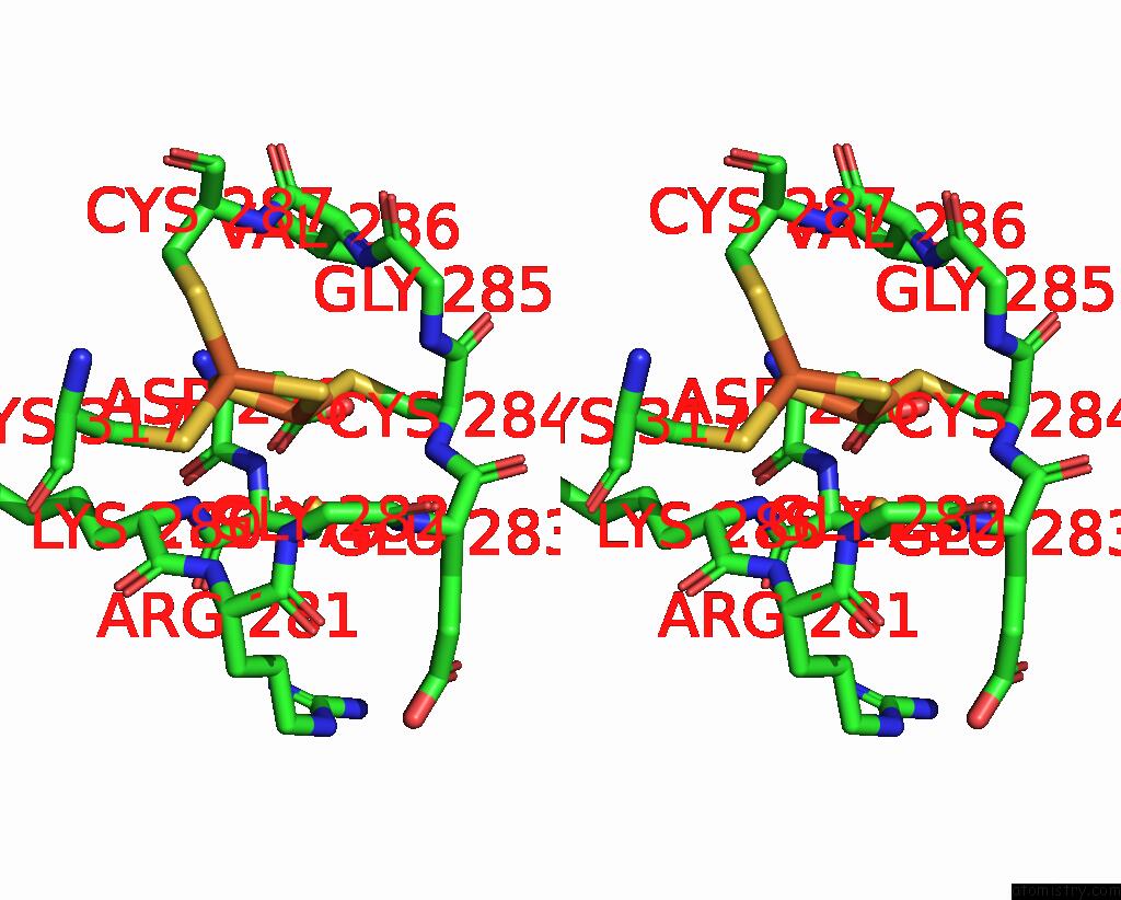
Stereo pair view
Mono view
Stereo pair view
A full contact list of Iron with other atoms in the Fe binding
site number 1 of Structure of A Bacteria Protein within 5.0Å range:
|
Iron binding site 2 out of 2 in 7ylr
Go back to
Iron binding site 2 out
of 2 in the Structure of A Bacteria Protein
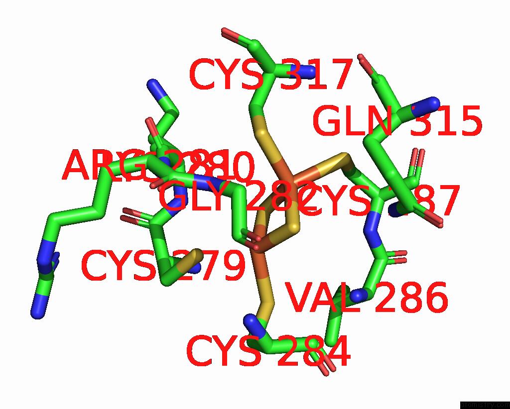
Mono view

Stereo pair view
Mono view
Stereo pair view
A full contact list of Iron with other atoms in the Fe binding
site number 2 of Structure of A Bacteria Protein within 5.0Å range:
|
Reference:
H.Zhang,
Y.J.Ma,
G.M.Yu,
X.Z.Li.
Structure of A Bacteria Protein To Be Published.
Page generated: Fri Aug 9 12:23:52 2024
Last articles
Fe in 7CNRFe in 7COH
Fe in 7CNT
Fe in 7CNS
Fe in 7CNQ
Fe in 7CNP
Fe in 7CN3
Fe in 7CKN
Fe in 7CL9
Fe in 7CL8