Iron »
PDB 7z0s-7zii »
7zif »
Iron in PDB 7zif: Crystal Structure of Human Tryptophan Hydroxylase 1 in Complex with Inhibitor Km-480
Enzymatic activity of Crystal Structure of Human Tryptophan Hydroxylase 1 in Complex with Inhibitor Km-480
All present enzymatic activity of Crystal Structure of Human Tryptophan Hydroxylase 1 in Complex with Inhibitor Km-480:
1.14.16.4;
1.14.16.4;
Protein crystallography data
The structure of Crystal Structure of Human Tryptophan Hydroxylase 1 in Complex with Inhibitor Km-480, PDB code: 7zif
was solved by
A.Schuetz,
N.Ziebart,
M.Weise,
K.Mallow,
J.Pfeifer,
M.Nazare,
E.Specker,
U.Heinemann,
with X-Ray Crystallography technique. A brief refinement statistics is given in the table below:
| Resolution Low / High (Å) | 41.29 / 1.87 |
| Space group | P 1 21 1 |
| Cell size a, b, c (Å), α, β, γ (°) | 47.021, 57.919, 59.416, 90, 97.65, 90 |
| R / Rfree (%) | 16 / 19.9 |
Iron Binding Sites:
The binding sites of Iron atom in the Crystal Structure of Human Tryptophan Hydroxylase 1 in Complex with Inhibitor Km-480
(pdb code 7zif). This binding sites where shown within
5.0 Angstroms radius around Iron atom.
In total only one binding site of Iron was determined in the Crystal Structure of Human Tryptophan Hydroxylase 1 in Complex with Inhibitor Km-480, PDB code: 7zif:
In total only one binding site of Iron was determined in the Crystal Structure of Human Tryptophan Hydroxylase 1 in Complex with Inhibitor Km-480, PDB code: 7zif:
Iron binding site 1 out of 1 in 7zif
Go back to
Iron binding site 1 out
of 1 in the Crystal Structure of Human Tryptophan Hydroxylase 1 in Complex with Inhibitor Km-480
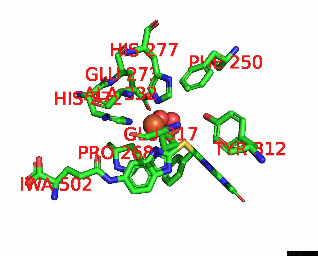
Mono view
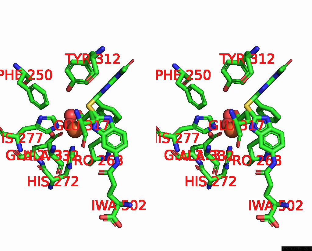
Stereo pair view
Mono view
Stereo pair view
A full contact list of Iron with other atoms in the Fe binding
site number 1 of Crystal Structure of Human Tryptophan Hydroxylase 1 in Complex with Inhibitor Km-480 within 5.0Å range:
|
Reference:
E.Specker,
S.Matthes,
R.Wesolowski,
A.Schutz,
M.Grohmann,
N.Alenina,
D.Pleimes,
K.Mallow,
M.Neuenschwander,
A.Gogolin,
M.Weise,
J.Pfeifer,
N.Ziebart,
U.Heinemann,
J.P.Von Kries,
M.Nazare,
M.Bader.
Structure-Based Design of Xanthine-Benzimidazole Derivatives As Novel and Potent Tryptophan Hydroxylase Inhibitors. J.Med.Chem. V. 65 11126 2022.
ISSN: ISSN 0022-2623
PubMed: 35921615
DOI: 10.1021/ACS.JMEDCHEM.2C00598
Page generated: Thu Aug 7 11:56:52 2025
ISSN: ISSN 0022-2623
PubMed: 35921615
DOI: 10.1021/ACS.JMEDCHEM.2C00598
Last articles
Xe in 2OQUXe in 2IE6
Xe in 2IC0
Xe in 2DKI
Xe in 2FIC
Xe in 2A7A
Xe in 2A7D
Xe in 2A9R
Xe in 2A7B
Xe in 1ZDM