Iron »
PDB 7zx5-8abf »
8a1h »
Iron in PDB 8a1h: Bacterial 6-4 Photolyase From Vibrio Cholerase
Protein crystallography data
The structure of Bacterial 6-4 Photolyase From Vibrio Cholerase, PDB code: 8a1h
was solved by
L.-O.Essen,
H.J.Emmerich,
with X-Ray Crystallography technique. A brief refinement statistics is given in the table below:
| Resolution Low / High (Å) | 34.59 / 1.65 |
| Space group | P 64 2 2 |
| Cell size a, b, c (Å), α, β, γ (°) | 199.687, 199.687, 76.996, 90, 90, 120 |
| R / Rfree (%) | 11.9 / 13.6 |
Other elements in 8a1h:
The structure of Bacterial 6-4 Photolyase From Vibrio Cholerase also contains other interesting chemical elements:
| Chlorine | (Cl) | 1 atom |
| Sodium | (Na) | 6 atoms |
Iron Binding Sites:
The binding sites of Iron atom in the Bacterial 6-4 Photolyase From Vibrio Cholerase
(pdb code 8a1h). This binding sites where shown within
5.0 Angstroms radius around Iron atom.
In total 4 binding sites of Iron where determined in the Bacterial 6-4 Photolyase From Vibrio Cholerase, PDB code: 8a1h:
Jump to Iron binding site number: 1; 2; 3; 4;
In total 4 binding sites of Iron where determined in the Bacterial 6-4 Photolyase From Vibrio Cholerase, PDB code: 8a1h:
Jump to Iron binding site number: 1; 2; 3; 4;
Iron binding site 1 out of 4 in 8a1h
Go back to
Iron binding site 1 out
of 4 in the Bacterial 6-4 Photolyase From Vibrio Cholerase
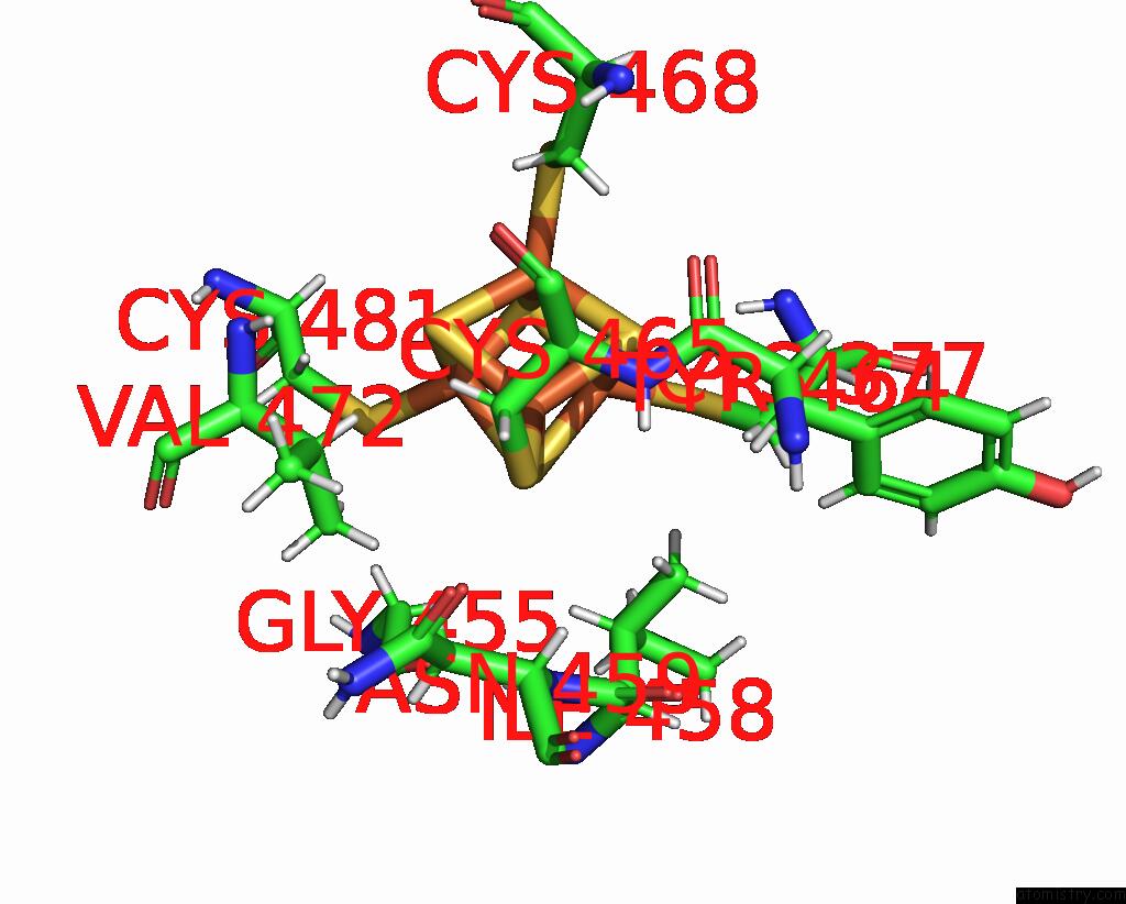
Mono view

Stereo pair view
Mono view
Stereo pair view
A full contact list of Iron with other atoms in the Fe binding
site number 1 of Bacterial 6-4 Photolyase From Vibrio Cholerase within 5.0Å range:
|
Iron binding site 2 out of 4 in 8a1h
Go back to
Iron binding site 2 out
of 4 in the Bacterial 6-4 Photolyase From Vibrio Cholerase
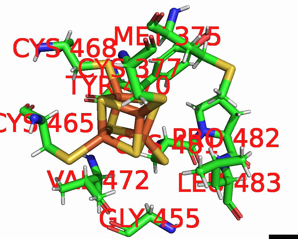
Mono view
Stereo pair view
Mono view
Stereo pair view
A full contact list of Iron with other atoms in the Fe binding
site number 2 of Bacterial 6-4 Photolyase From Vibrio Cholerase within 5.0Å range:
|
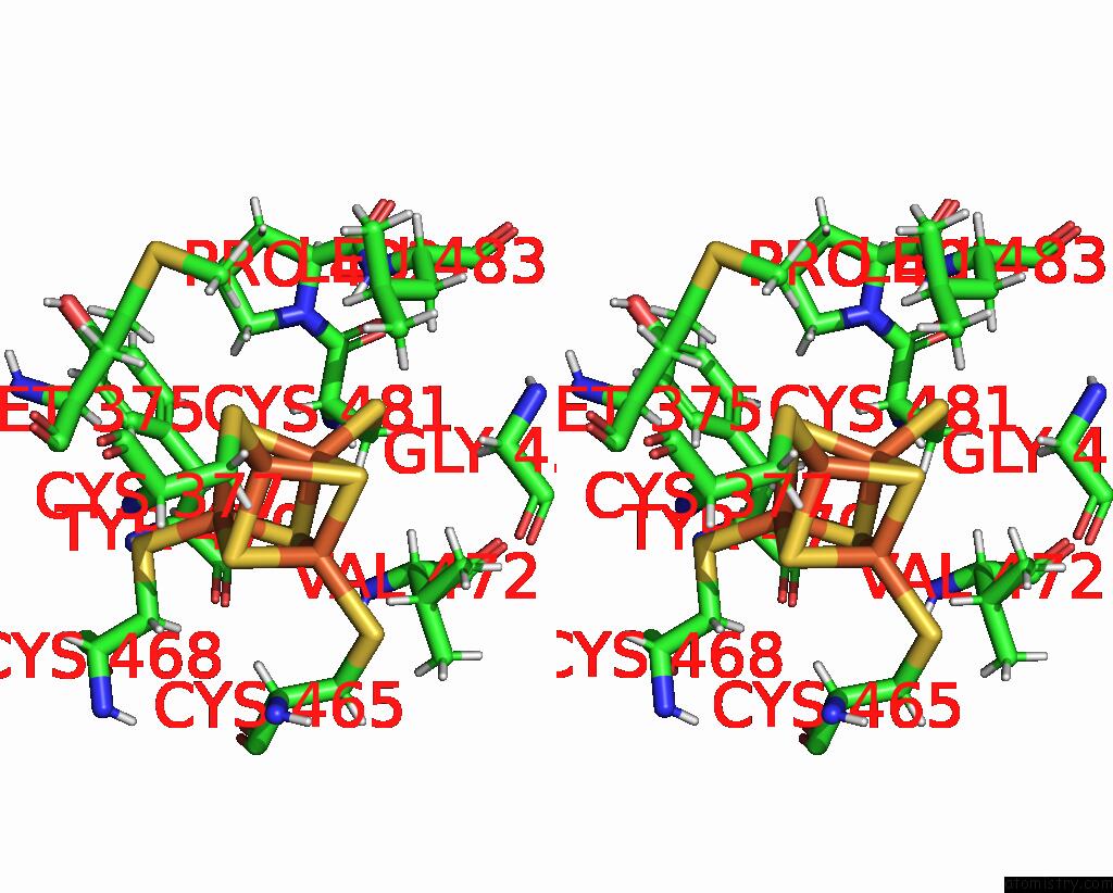
Iron binding site 3 out of 4 in 8a1h
Go back to
Iron binding site 3 out
of 4 in the Bacterial 6-4 Photolyase From Vibrio Cholerase

Mono view

Stereo pair view
Mono view
Stereo pair view
A full contact list of Iron with other atoms in the Fe binding
site number 3 of Bacterial 6-4 Photolyase From Vibrio Cholerase within 5.0Å range:
|
Iron binding site 4 out of 4 in 8a1h
Go back to
Iron binding site 4 out
of 4 in the Bacterial 6-4 Photolyase From Vibrio Cholerase

Mono view
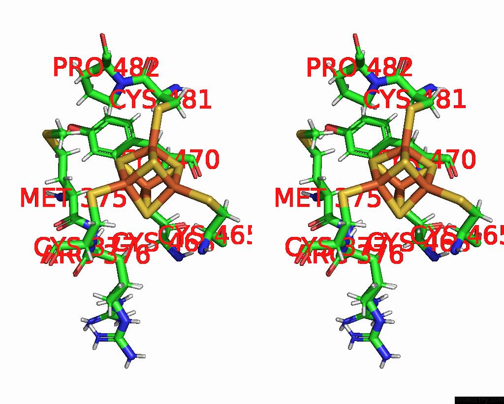
Stereo pair view
Mono view
Stereo pair view
A full contact list of Iron with other atoms in the Fe binding
site number 4 of Bacterial 6-4 Photolyase From Vibrio Cholerase within 5.0Å range:
|
Reference:
H.J.Emmerich,
L.Schneider,
L.O.Essen.
Structural and Functional Analysis of A Prokaryotic (6-4) Photolyase From the Aquatic Pathogen Vibrio Cholerae. Photochem.Photobiol. 2023.
ISSN: ISSN 0031-8655
PubMed: 36692077
DOI: 10.1111/PHP.13783
Page generated: Fri Aug 9 16:02:04 2024
ISSN: ISSN 0031-8655
PubMed: 36692077
DOI: 10.1111/PHP.13783
Last articles
Zn in 9MJ5Zn in 9HNW
Zn in 9G0L
Zn in 9FNE
Zn in 9DZN
Zn in 9E0I
Zn in 9D32
Zn in 9DAK
Zn in 8ZXC
Zn in 8ZUF