Iron »
PDB 8abi-8asi »
8ad0 »
Iron in PDB 8ad0: X-Ray Structure of Na+-Nqr From Vibrio Cholerae in Different Conformation at 3.1 A
Enzymatic activity of X-Ray Structure of Na+-Nqr From Vibrio Cholerae in Different Conformation at 3.1 A
All present enzymatic activity of X-Ray Structure of Na+-Nqr From Vibrio Cholerae in Different Conformation at 3.1 A:
7.2.1.1;
7.2.1.1;
Protein crystallography data
The structure of X-Ray Structure of Na+-Nqr From Vibrio Cholerae in Different Conformation at 3.1 A, PDB code: 8ad0
was solved by
G.Fritz,
with X-Ray Crystallography technique. A brief refinement statistics is given in the table below:
| Resolution Low / High (Å) | 48.12 / 3.11 |
| Space group | P 1 21 1 |
| Cell size a, b, c (Å), α, β, γ (°) | 89.31, 142.15, 105.93, 90, 109.83, 90 |
| R / Rfree (%) | 24.9 / 28.8 |
Other elements in 8ad0:
The structure of X-Ray Structure of Na+-Nqr From Vibrio Cholerae in Different Conformation at 3.1 A also contains other interesting chemical elements:
| Bromine | (Br) | 1 atom |
| Sodium | (Na) | 1 atom |
| Potassium | (K) | 1 atom |
Iron Binding Sites:
The binding sites of Iron atom in the X-Ray Structure of Na+-Nqr From Vibrio Cholerae in Different Conformation at 3.1 A
(pdb code 8ad0). This binding sites where shown within
5.0 Angstroms radius around Iron atom.
In total 4 binding sites of Iron where determined in the X-Ray Structure of Na+-Nqr From Vibrio Cholerae in Different Conformation at 3.1 A, PDB code: 8ad0:
Jump to Iron binding site number: 1; 2; 3; 4;
In total 4 binding sites of Iron where determined in the X-Ray Structure of Na+-Nqr From Vibrio Cholerae in Different Conformation at 3.1 A, PDB code: 8ad0:
Jump to Iron binding site number: 1; 2; 3; 4;
Iron binding site 1 out of 4 in 8ad0
Go back to
Iron binding site 1 out
of 4 in the X-Ray Structure of Na+-Nqr From Vibrio Cholerae in Different Conformation at 3.1 A
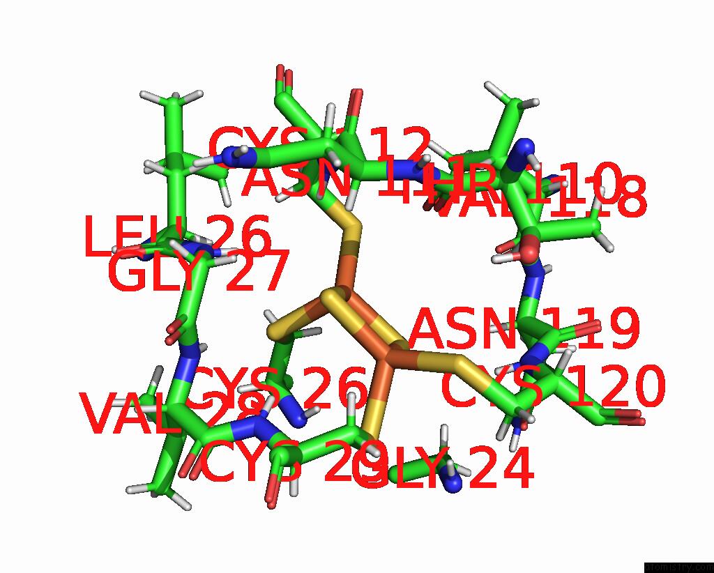
Mono view
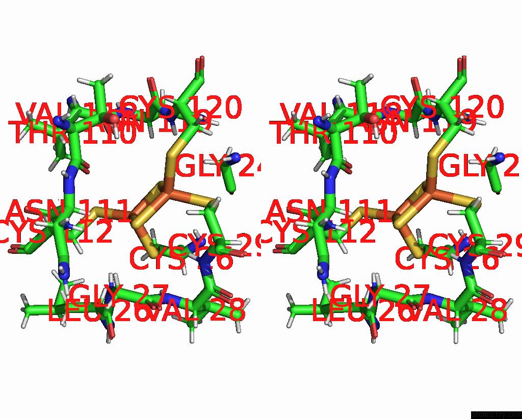
Stereo pair view
Mono view
Stereo pair view
A full contact list of Iron with other atoms in the Fe binding
site number 1 of X-Ray Structure of Na+-Nqr From Vibrio Cholerae in Different Conformation at 3.1 A within 5.0Å range:
|
Iron binding site 2 out of 4 in 8ad0
Go back to
Iron binding site 2 out
of 4 in the X-Ray Structure of Na+-Nqr From Vibrio Cholerae in Different Conformation at 3.1 A

Mono view

Stereo pair view
Mono view
Stereo pair view
A full contact list of Iron with other atoms in the Fe binding
site number 2 of X-Ray Structure of Na+-Nqr From Vibrio Cholerae in Different Conformation at 3.1 A within 5.0Å range:
|
Iron binding site 3 out of 4 in 8ad0
Go back to
Iron binding site 3 out
of 4 in the X-Ray Structure of Na+-Nqr From Vibrio Cholerae in Different Conformation at 3.1 A
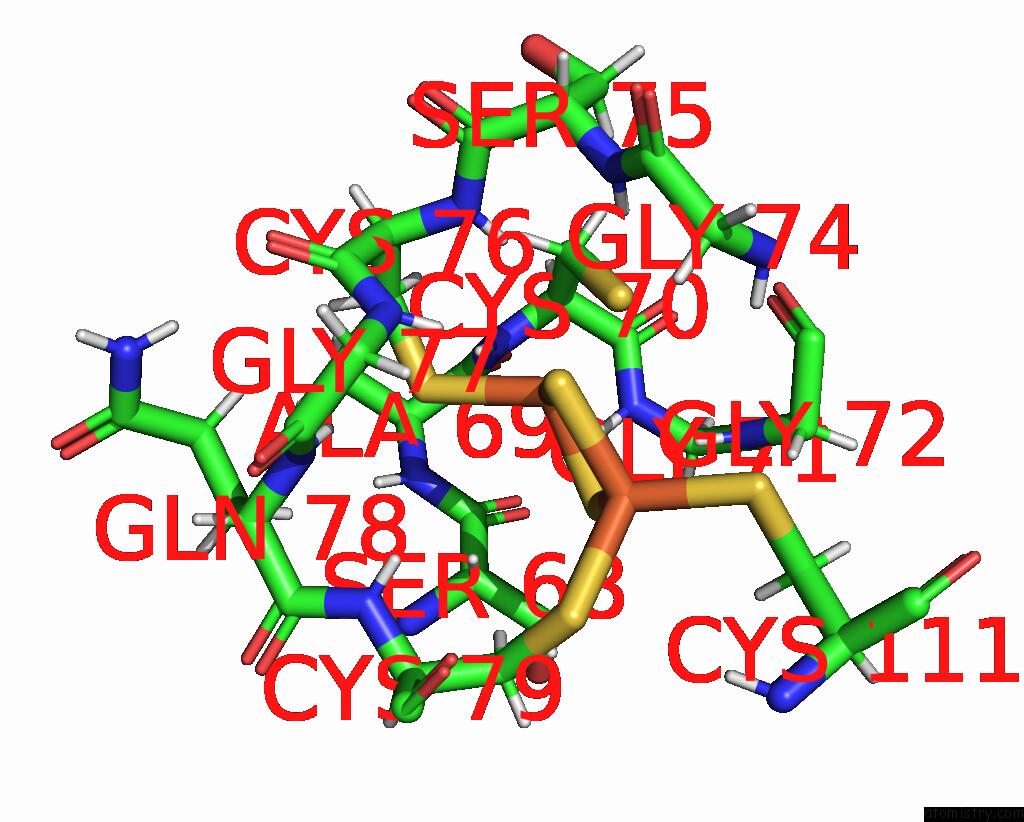
Mono view

Stereo pair view
Mono view
Stereo pair view
A full contact list of Iron with other atoms in the Fe binding
site number 3 of X-Ray Structure of Na+-Nqr From Vibrio Cholerae in Different Conformation at 3.1 A within 5.0Å range:
|
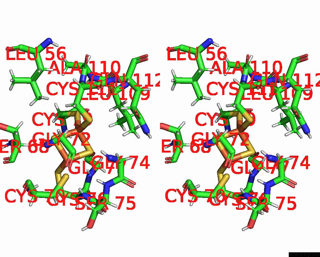
Iron binding site 4 out of 4 in 8ad0
Go back to
Iron binding site 4 out
of 4 in the X-Ray Structure of Na+-Nqr From Vibrio Cholerae in Different Conformation at 3.1 A

Mono view
Stereo pair view
Mono view
Stereo pair view
A full contact list of Iron with other atoms in the Fe binding
site number 4 of X-Ray Structure of Na+-Nqr From Vibrio Cholerae in Different Conformation at 3.1 A within 5.0Å range:
|
Reference:
G.Fritz,
G.Fritz.
N/A N/A.
ISSN: ESSN 1545-9985
Page generated: Thu Aug 7 12:57:16 2025
ISSN: ESSN 1545-9985
Last articles
I in 5IO8I in 5IJS
I in 5IJW
I in 5GZH
I in 5IE1
I in 5I3W
I in 5HOW
I in 5GLS
I in 5I3V
I in 5HPP