Iron »
PDB 8euh-8f9n »
8ewl »
Iron in PDB 8ewl: Crystal Structure of CYP3A4 Bound to An Inhibitor
Enzymatic activity of Crystal Structure of CYP3A4 Bound to An Inhibitor
All present enzymatic activity of Crystal Structure of CYP3A4 Bound to An Inhibitor:
1.14.14.1; 1.14.14.55; 1.14.14.56; 1.14.14.73;
1.14.14.1; 1.14.14.55; 1.14.14.56; 1.14.14.73;
Protein crystallography data
The structure of Crystal Structure of CYP3A4 Bound to An Inhibitor, PDB code: 8ewl
was solved by
I.F.Sevrioukova,
with X-Ray Crystallography technique. A brief refinement statistics is given in the table below:
| Resolution Low / High (Å) | 44.42 / 2.35 |
| Space group | I 2 2 2 |
| Cell size a, b, c (Å), α, β, γ (°) | 77.079, 100.819, 129.099, 90, 90, 90 |
| R / Rfree (%) | 21.6 / 26.5 |
Other elements in 8ewl:
The structure of Crystal Structure of CYP3A4 Bound to An Inhibitor also contains other interesting chemical elements:
| Iridium | (Ir) | 1 atom |
Iron Binding Sites:
The binding sites of Iron atom in the Crystal Structure of CYP3A4 Bound to An Inhibitor
(pdb code 8ewl). This binding sites where shown within
5.0 Angstroms radius around Iron atom.
In total only one binding site of Iron was determined in the Crystal Structure of CYP3A4 Bound to An Inhibitor, PDB code: 8ewl:
In total only one binding site of Iron was determined in the Crystal Structure of CYP3A4 Bound to An Inhibitor, PDB code: 8ewl:
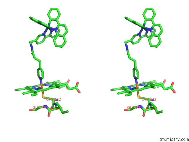
Iron binding site 1 out of 1 in 8ewl
Go back to
Iron binding site 1 out
of 1 in the Crystal Structure of CYP3A4 Bound to An Inhibitor
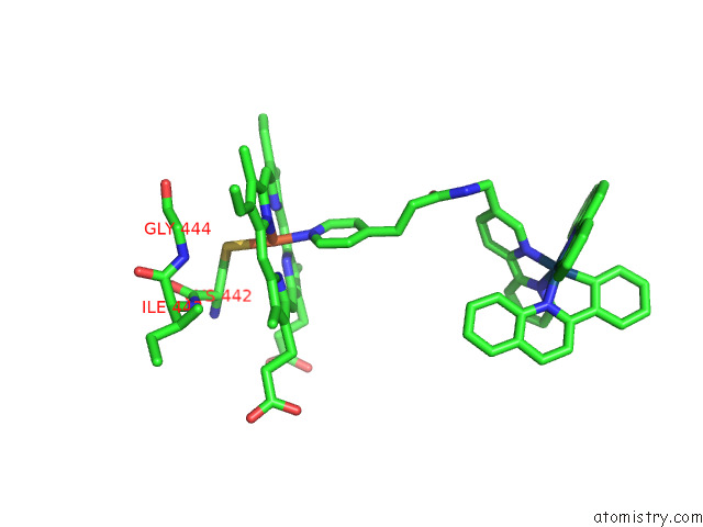
Mono view
Stereo pair view
Mono view
Stereo pair view
A full contact list of Iron with other atoms in the Fe binding
site number 1 of Crystal Structure of CYP3A4 Bound to An Inhibitor within 5.0Å range:
|
Reference:
M.Denison,
J.Ahrens,
M.Dunbar,
H.Warmahaye,
A.Majeed,
C.Turro,
T.Kocarek,
I.F.Sevrioukova,
J.Kodanko.
Dynamic Ir(III) Photosensors For the Major Human Drug Metabolizing Enzyme Cytochrome P450 3A4 To Be Published.
Page generated: Thu Aug 7 16:44:47 2025
Last articles
Mg in 1W25Mg in 1W23
Mg in 1W1Z
Mg in 1W1W
Mg in 1W0N
Mg in 1W0K
Mg in 1W0J
Mg in 1W0H
Mg in 1VZM
Mg in 1VZK