Iron »
PDB 8fay-8fib »
8fay »
Iron in PDB 8fay: Human Mutyh Adenine Glycosylase Bound to Dna Containing A Transition State Analog (1N) Paired with D(8-Oxo-G)
Enzymatic activity of Human Mutyh Adenine Glycosylase Bound to Dna Containing A Transition State Analog (1N) Paired with D(8-Oxo-G)
All present enzymatic activity of Human Mutyh Adenine Glycosylase Bound to Dna Containing A Transition State Analog (1N) Paired with D(8-Oxo-G):
3.2.2.31;
3.2.2.31;
Protein crystallography data
The structure of Human Mutyh Adenine Glycosylase Bound to Dna Containing A Transition State Analog (1N) Paired with D(8-Oxo-G), PDB code: 8fay
was solved by
C.H.Trasvina-Arenas,
W.J.Lin,
M.Demir,
A.J.Fisher,
S.S.David,
M.P.Horvath,
with X-Ray Crystallography technique. A brief refinement statistics is given in the table below:
| Resolution Low / High (Å) | 60.28 / 1.91 |
| Space group | P 21 21 21 |
| Cell size a, b, c (Å), α, β, γ (°) | 87.686, 116.422, 118.378, 90, 90, 90 |
| R / Rfree (%) | 18.1 / 20.9 |
Iron Binding Sites:
The binding sites of Iron atom in the Human Mutyh Adenine Glycosylase Bound to Dna Containing A Transition State Analog (1N) Paired with D(8-Oxo-G)
(pdb code 8fay). This binding sites where shown within
5.0 Angstroms radius around Iron atom.
In total 8 binding sites of Iron where determined in the Human Mutyh Adenine Glycosylase Bound to Dna Containing A Transition State Analog (1N) Paired with D(8-Oxo-G), PDB code: 8fay:
Jump to Iron binding site number: 1; 2; 3; 4; 5; 6; 7; 8;
In total 8 binding sites of Iron where determined in the Human Mutyh Adenine Glycosylase Bound to Dna Containing A Transition State Analog (1N) Paired with D(8-Oxo-G), PDB code: 8fay:
Jump to Iron binding site number: 1; 2; 3; 4; 5; 6; 7; 8;
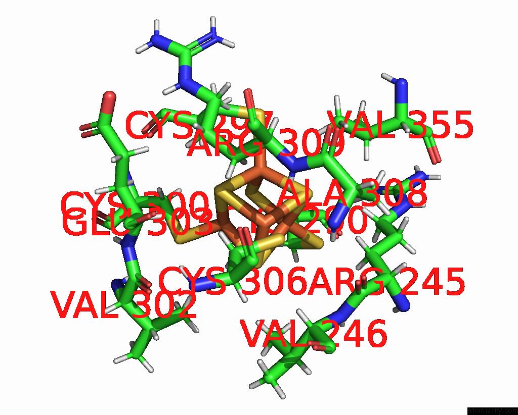
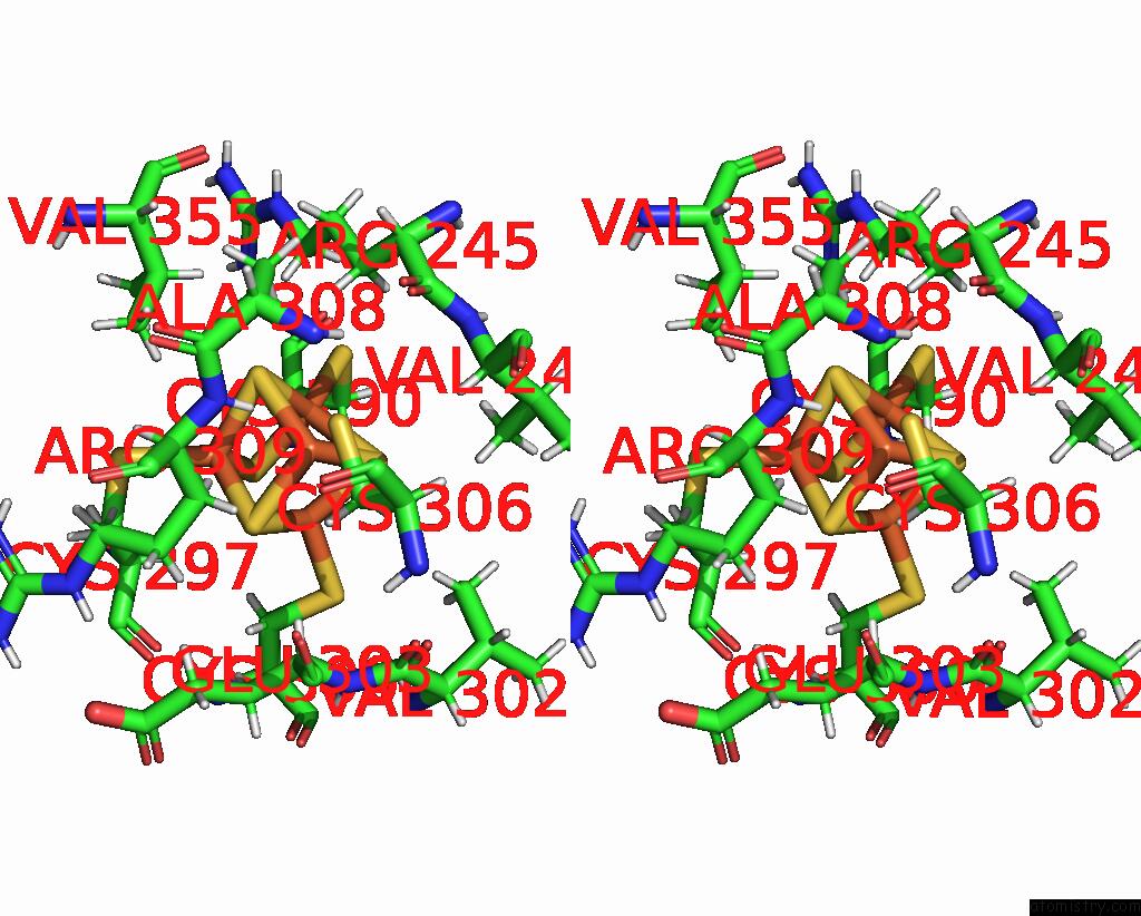
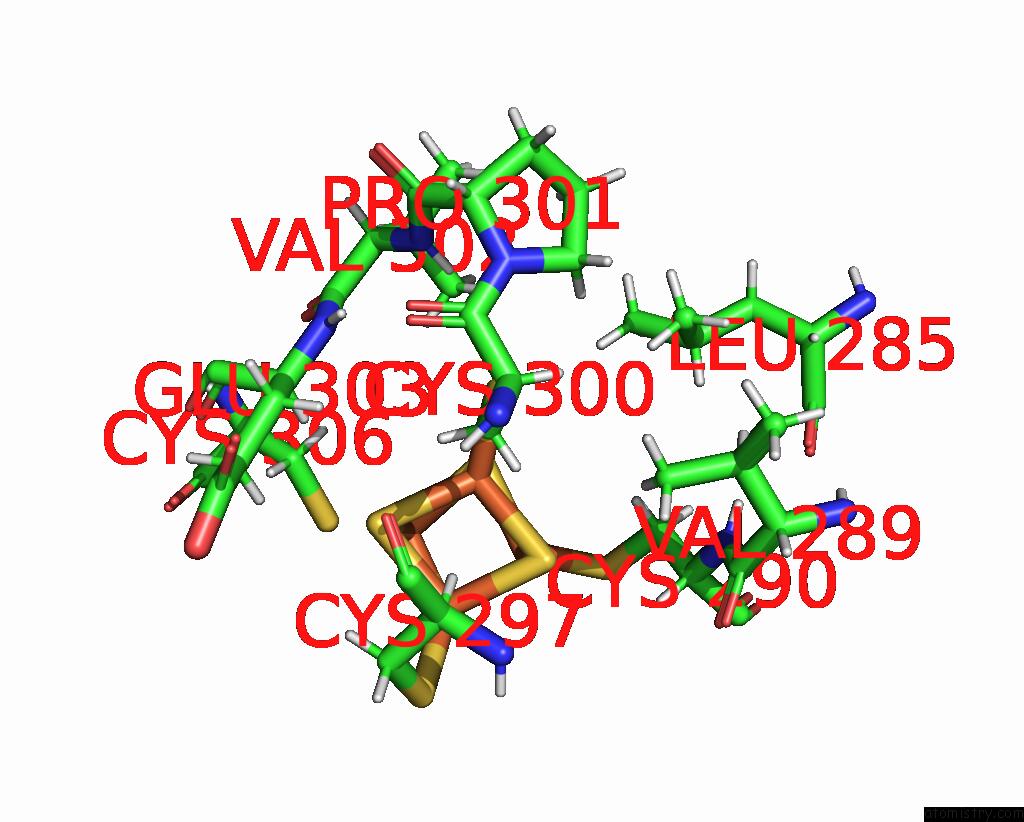
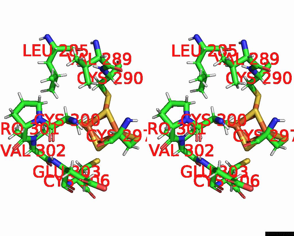
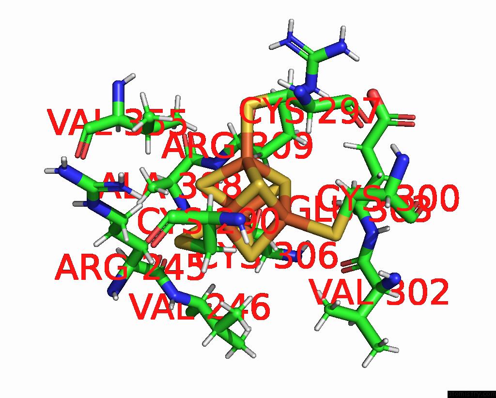
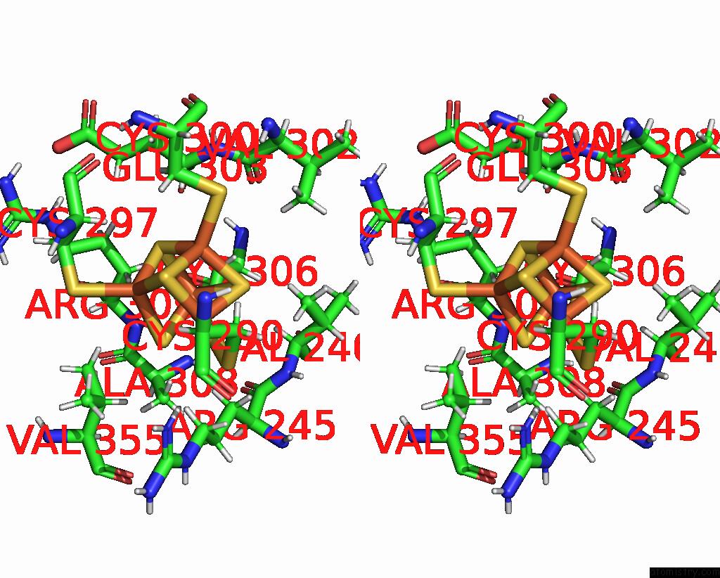
Iron binding site 1 out of 8 in 8fay
Go back to
Iron binding site 1 out
of 8 in the Human Mutyh Adenine Glycosylase Bound to Dna Containing A Transition State Analog (1N) Paired with D(8-Oxo-G)
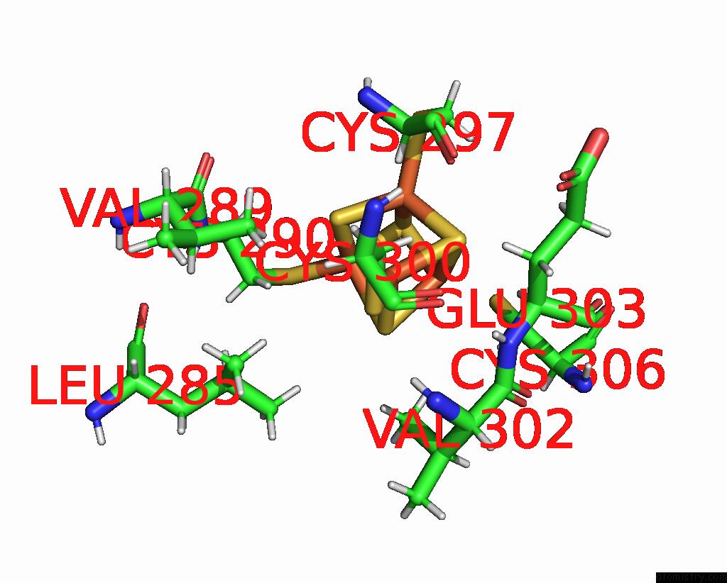
Mono view
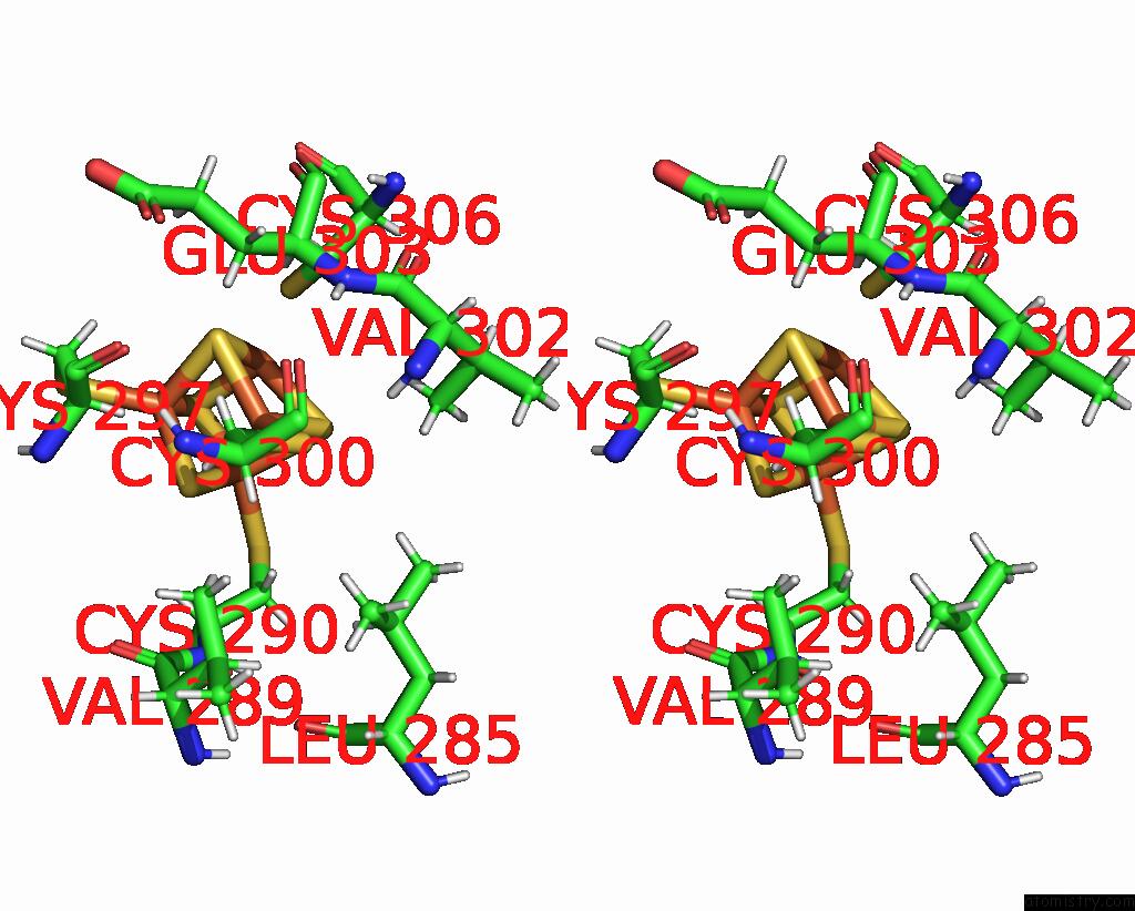
Stereo pair view
Mono view
Stereo pair view
|
|
A full contact list of Iron with other atoms in the Fe binding
site number 1 of Human Mutyh Adenine Glycosylase Bound to Dna Containing A Transition State Analog (1N) Paired with D(8-Oxo-G) within 5.0Å range:
|
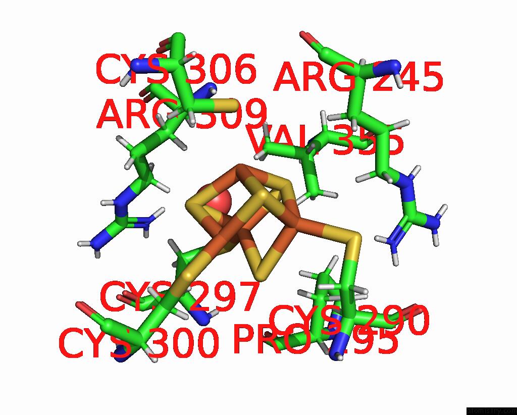
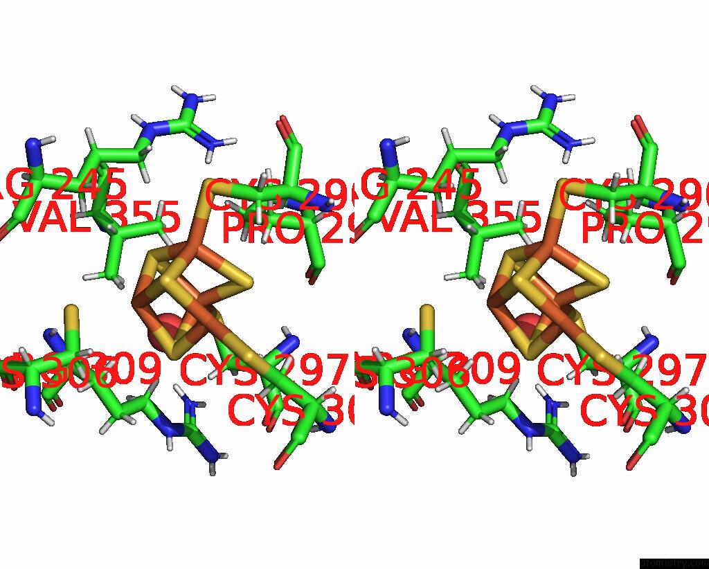
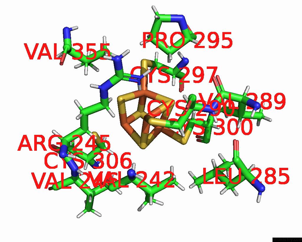
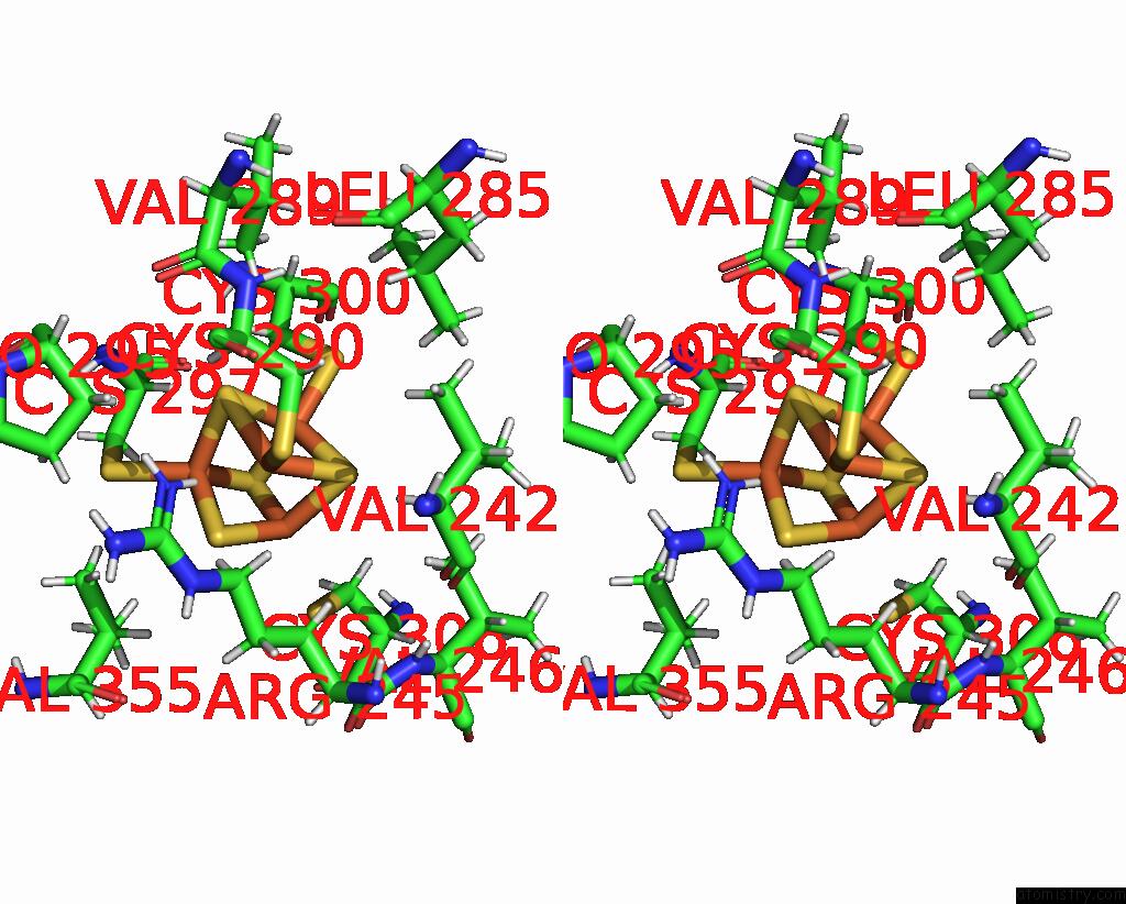
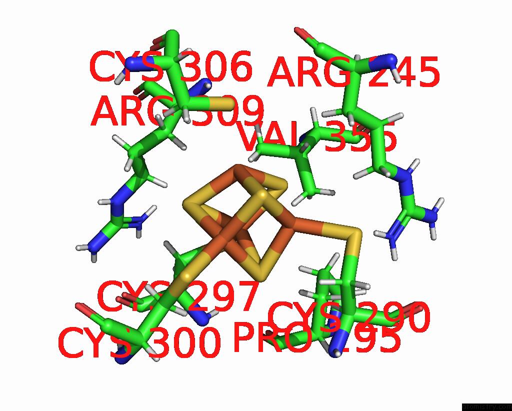
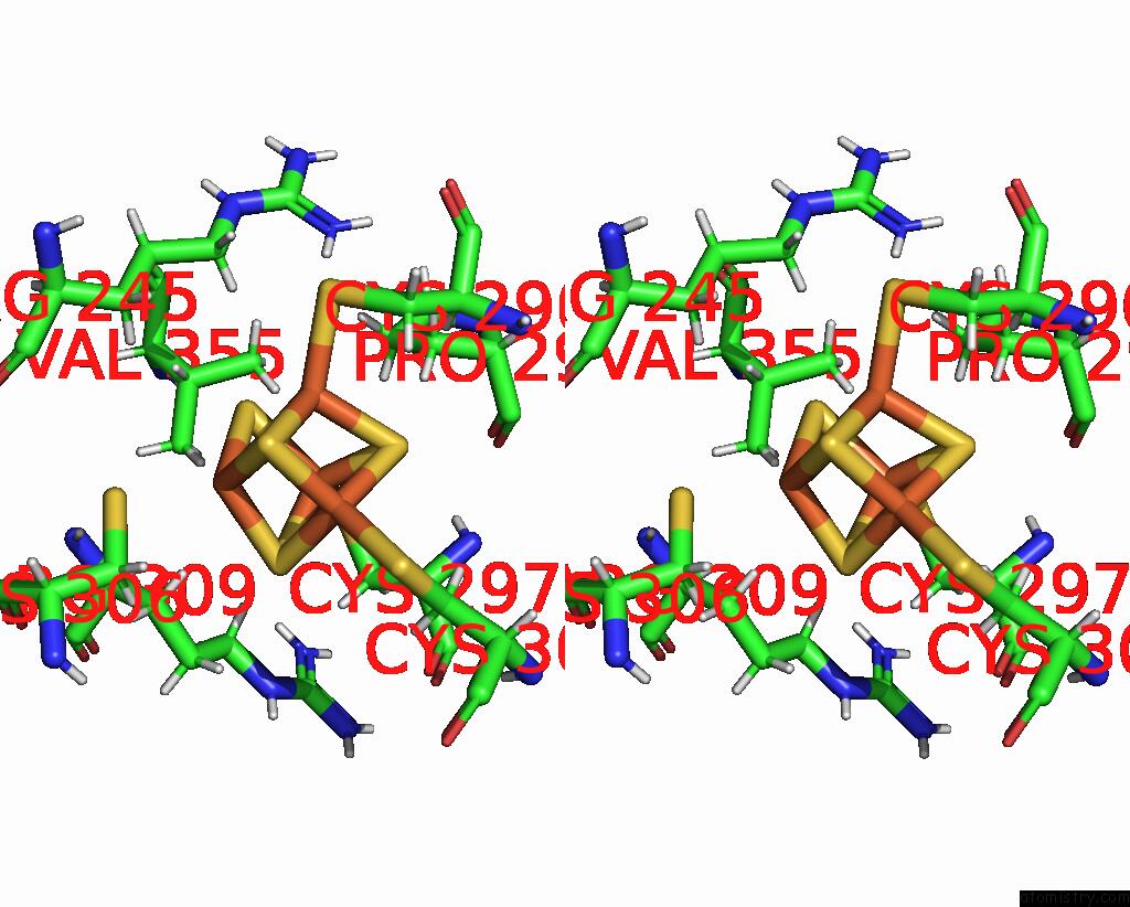
Iron binding site 2 out of 8 in 8fay
Go back to
Iron binding site 2 out
of 8 in the Human Mutyh Adenine Glycosylase Bound to Dna Containing A Transition State Analog (1N) Paired with D(8-Oxo-G)
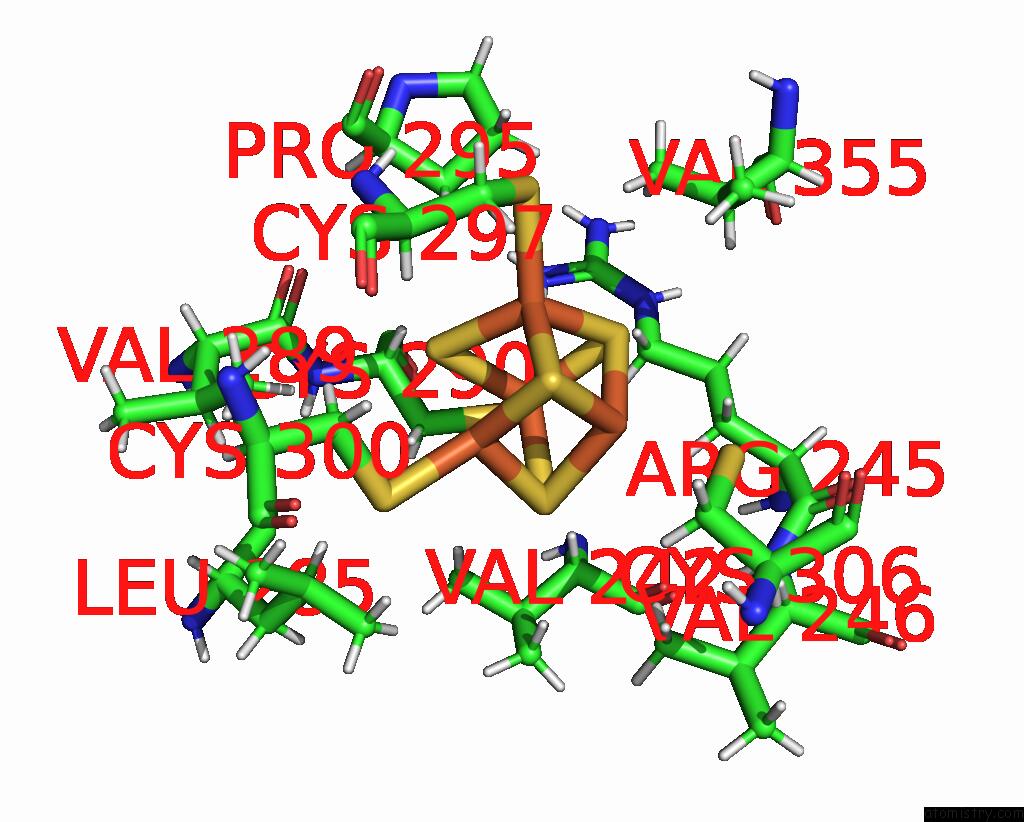
Mono view
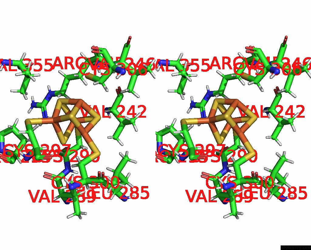
Stereo pair view
Mono view
Stereo pair view
|
|
A full contact list of Iron with other atoms in the Fe binding
site number 2 of Human Mutyh Adenine Glycosylase Bound to Dna Containing A Transition State Analog (1N) Paired with D(8-Oxo-G) within 5.0Å range:
|
Iron binding site 3 out of 8 in 8fay
Go back to
Iron binding site 3 out
of 8 in the Human Mutyh Adenine Glycosylase Bound to Dna Containing A Transition State Analog (1N) Paired with D(8-Oxo-G)
Mono view
Stereo pair view
Mono view
Stereo pair view
|
|
A full contact list of Iron with other atoms in the Fe binding
site number 3 of Human Mutyh Adenine Glycosylase Bound to Dna Containing A Transition State Analog (1N) Paired with D(8-Oxo-G) within 5.0Å range:
|
Iron binding site 4 out of 8 in 8fay
Go back to
Iron binding site 4 out
of 8 in the Human Mutyh Adenine Glycosylase Bound to Dna Containing A Transition State Analog (1N) Paired with D(8-Oxo-G)
Mono view
Stereo pair view
Mono view
Stereo pair view
|
|
A full contact list of Iron with other atoms in the Fe binding
site number 4 of Human Mutyh Adenine Glycosylase Bound to Dna Containing A Transition State Analog (1N) Paired with D(8-Oxo-G) within 5.0Å range:
|
Iron binding site 5 out of 8 in 8fay
Go back to
Iron binding site 5 out
of 8 in the Human Mutyh Adenine Glycosylase Bound to Dna Containing A Transition State Analog (1N) Paired with D(8-Oxo-G)
Mono view
Stereo pair view
Mono view
Stereo pair view
|
|
A full contact list of Iron with other atoms in the Fe binding
site number 5 of Human Mutyh Adenine Glycosylase Bound to Dna Containing A Transition State Analog (1N) Paired with D(8-Oxo-G) within 5.0Å range:
|
Iron binding site 6 out of 8 in 8fay
Go back to
Iron binding site 6 out
of 8 in the Human Mutyh Adenine Glycosylase Bound to Dna Containing A Transition State Analog (1N) Paired with D(8-Oxo-G)
Mono view
Stereo pair view
Mono view
Stereo pair view
|
|
A full contact list of Iron with other atoms in the Fe binding
site number 6 of Human Mutyh Adenine Glycosylase Bound to Dna Containing A Transition State Analog (1N) Paired with D(8-Oxo-G) within 5.0Å range:
|
Iron binding site 7 out of 8 in 8fay
Go back to
Iron binding site 7 out
of 8 in the Human Mutyh Adenine Glycosylase Bound to Dna Containing A Transition State Analog (1N) Paired with D(8-Oxo-G)
Mono view
Stereo pair view
Mono view
Stereo pair view
|
|
A full contact list of Iron with other atoms in the Fe binding
site number 7 of Human Mutyh Adenine Glycosylase Bound to Dna Containing A Transition State Analog (1N) Paired with D(8-Oxo-G) within 5.0Å range:
|
Iron binding site 8 out of 8 in 8fay
Go back to
Iron binding site 8 out
of 8 in the Human Mutyh Adenine Glycosylase Bound to Dna Containing A Transition State Analog (1N) Paired with D(8-Oxo-G)
Mono view
Stereo pair view
Mono view
Stereo pair view
|
|
A full contact list of Iron with other atoms in the Fe binding
site number 8 of Human Mutyh Adenine Glycosylase Bound to Dna Containing A Transition State Analog (1N) Paired with D(8-Oxo-G) within 5.0Å range:
|
Reference:
C.H.Trasvina-Arenas,
C.H.Trasvina-Arenas,
W.J.Lin,
M.Demir,
A.J.Fisher,
S.S.David,
M.P.Horvath.
N/A N/A.
Page generated: Thu Aug 7 17:08:47 2025
Last articles
Mg in 9IRPMg in 9IRE
Mg in 9IQY
Mg in 9IQW
Mg in 9IQX
Mg in 9IO5
Mg in 9IQG
Mg in 9IMR
Mg in 9IMQ
Mg in 9IM2