Iron »
PDB 8fic-8gcq »
8fjo »
Iron in PDB 8fjo: X-Ray Crystal Structure of CYP124A1 From Mycobacterium Marinum in Complex with Farnesyl Acetate
Protein crystallography data
The structure of X-Ray Crystal Structure of CYP124A1 From Mycobacterium Marinum in Complex with Farnesyl Acetate, PDB code: 8fjo
was solved by
A.Ghith,
J.B.Bruning,
S.G.Bell,
with X-Ray Crystallography technique. A brief refinement statistics is given in the table below:
| Resolution Low / High (Å) | 45.92 / 1.69 |
| Space group | C 1 2 1 |
| Cell size a, b, c (Å), α, β, γ (°) | 97.511, 71.635, 64.624, 90, 109.64, 90 |
| R / Rfree (%) | 17.7 / 21.1 |
Iron Binding Sites:
The binding sites of Iron atom in the X-Ray Crystal Structure of CYP124A1 From Mycobacterium Marinum in Complex with Farnesyl Acetate
(pdb code 8fjo). This binding sites where shown within
5.0 Angstroms radius around Iron atom.
In total only one binding site of Iron was determined in the X-Ray Crystal Structure of CYP124A1 From Mycobacterium Marinum in Complex with Farnesyl Acetate, PDB code: 8fjo:
In total only one binding site of Iron was determined in the X-Ray Crystal Structure of CYP124A1 From Mycobacterium Marinum in Complex with Farnesyl Acetate, PDB code: 8fjo:
Iron binding site 1 out of 1 in 8fjo
Go back to
Iron binding site 1 out
of 1 in the X-Ray Crystal Structure of CYP124A1 From Mycobacterium Marinum in Complex with Farnesyl Acetate
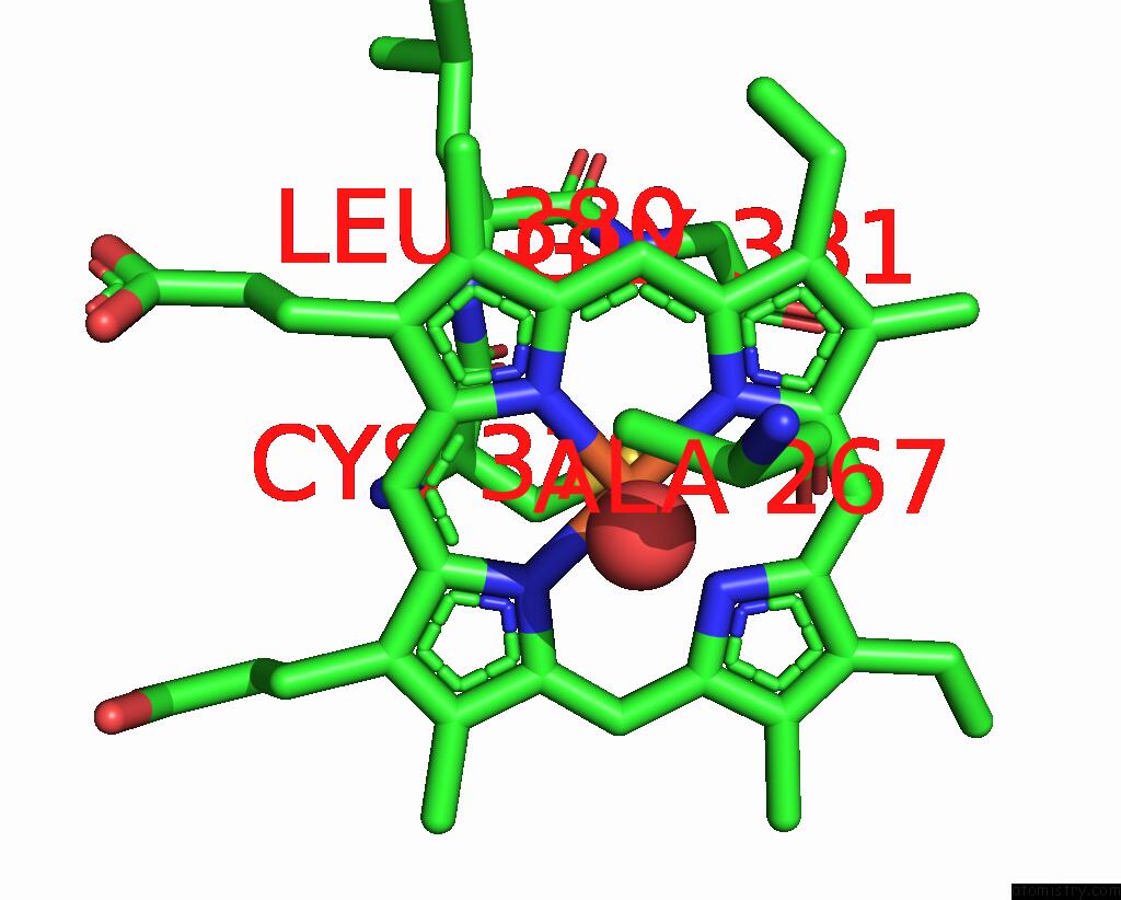
Mono view
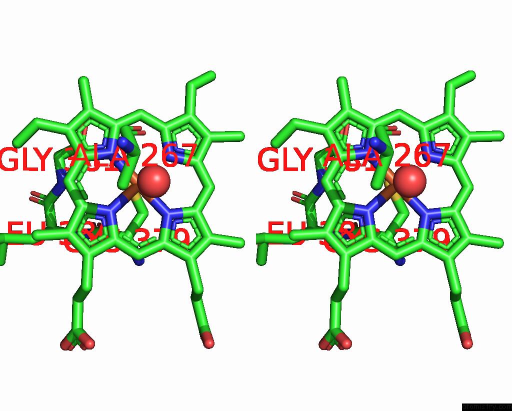
Stereo pair view
Mono view
Stereo pair view
|
|
A full contact list of Iron with other atoms in the Fe binding
site number 1 of X-Ray Crystal Structure of CYP124A1 From Mycobacterium Marinum in Complex with Farnesyl Acetate within 5.0Å range:
|
Reference:
A.Ghith,
J.B.Bruning,
S.G.Bell.
The Catalytic Activity and Structure of the Lipid Metabolizing CYP124 Cytochrome P450 Enzyme From Mycobacterium Marinum. Arch.Biochem.Biophys. V. 737 09554 2023.
ISSN: ESSN 1096-0384
PubMed: 36842492
DOI: 10.1016/J.ABB.2023.109554
Page generated: Thu Aug 7 17:21:59 2025
ISSN: ESSN 1096-0384
PubMed: 36842492
DOI: 10.1016/J.ABB.2023.109554
Last articles
Pt in 3IWLPt in 3ESB
Pt in 3EF3
Pt in 2XN0
Pt in 3AZ7
Pt in 3AZ5
Pt in 3CO3
Pt in 2ZZB
Pt in 2Z8Z
Pt in 2XTH