Iron »
PDB 8hcr-8ib5 »
8hdd »
Iron in PDB 8hdd: Complex Structure of Catalytic, Small, and A Partial Electron Transfer Subunits From Burkholderia Cepacia Fad Glucose Dehydrogenase
Enzymatic activity of Complex Structure of Catalytic, Small, and A Partial Electron Transfer Subunits From Burkholderia Cepacia Fad Glucose Dehydrogenase
All present enzymatic activity of Complex Structure of Catalytic, Small, and A Partial Electron Transfer Subunits From Burkholderia Cepacia Fad Glucose Dehydrogenase:
1.1.5.9;
1.1.5.9;
Protein crystallography data
The structure of Complex Structure of Catalytic, Small, and A Partial Electron Transfer Subunits From Burkholderia Cepacia Fad Glucose Dehydrogenase, PDB code: 8hdd
was solved by
H.Yoshida,
K.Sode,
with X-Ray Crystallography technique. A brief refinement statistics is given in the table below:
| Resolution Low / High (Å) | 49.88 / 3.00 |
| Space group | P 21 21 2 |
| Cell size a, b, c (Å), α, β, γ (°) | 204.015, 71.797, 114.221, 90, 90, 90 |
| R / Rfree (%) | 27.5 / 32.3 |
Iron Binding Sites:
The binding sites of Iron atom in the Complex Structure of Catalytic, Small, and A Partial Electron Transfer Subunits From Burkholderia Cepacia Fad Glucose Dehydrogenase
(pdb code 8hdd). This binding sites where shown within
5.0 Angstroms radius around Iron atom.
In total 4 binding sites of Iron where determined in the Complex Structure of Catalytic, Small, and A Partial Electron Transfer Subunits From Burkholderia Cepacia Fad Glucose Dehydrogenase, PDB code: 8hdd:
Jump to Iron binding site number: 1; 2; 3; 4;
In total 4 binding sites of Iron where determined in the Complex Structure of Catalytic, Small, and A Partial Electron Transfer Subunits From Burkholderia Cepacia Fad Glucose Dehydrogenase, PDB code: 8hdd:
Jump to Iron binding site number: 1; 2; 3; 4;
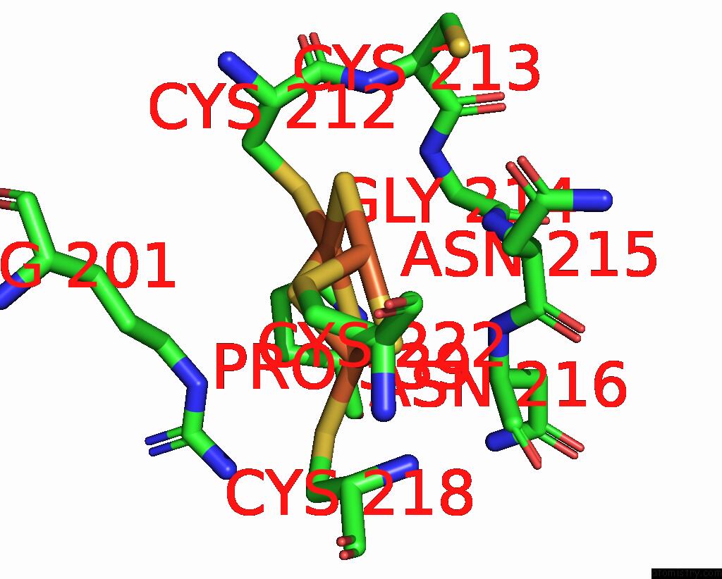
Iron binding site 1 out of 4 in 8hdd
Go back to
Iron binding site 1 out
of 4 in the Complex Structure of Catalytic, Small, and A Partial Electron Transfer Subunits From Burkholderia Cepacia Fad Glucose Dehydrogenase
Mono view

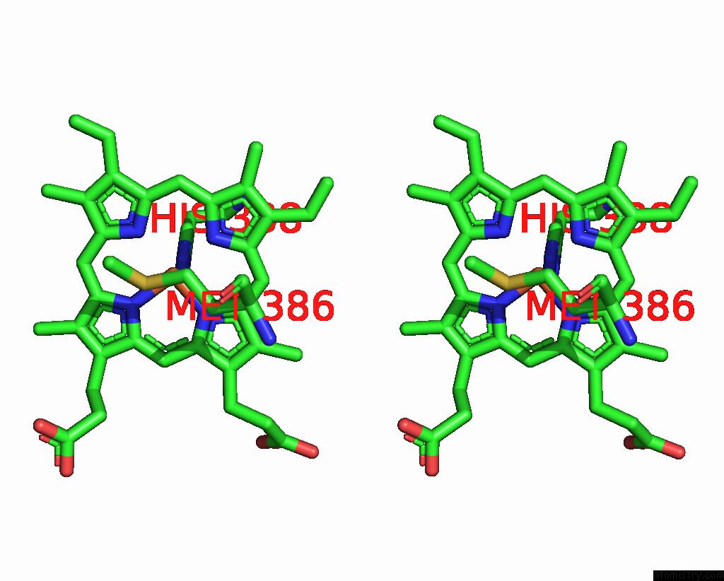
Stereo pair view
Mono view
Stereo pair view
|
|
A full contact list of Iron with other atoms in the Fe binding
site number 1 of Complex Structure of Catalytic, Small, and A Partial Electron Transfer Subunits From Burkholderia Cepacia Fad Glucose Dehydrogenase within 5.0Å range:
|
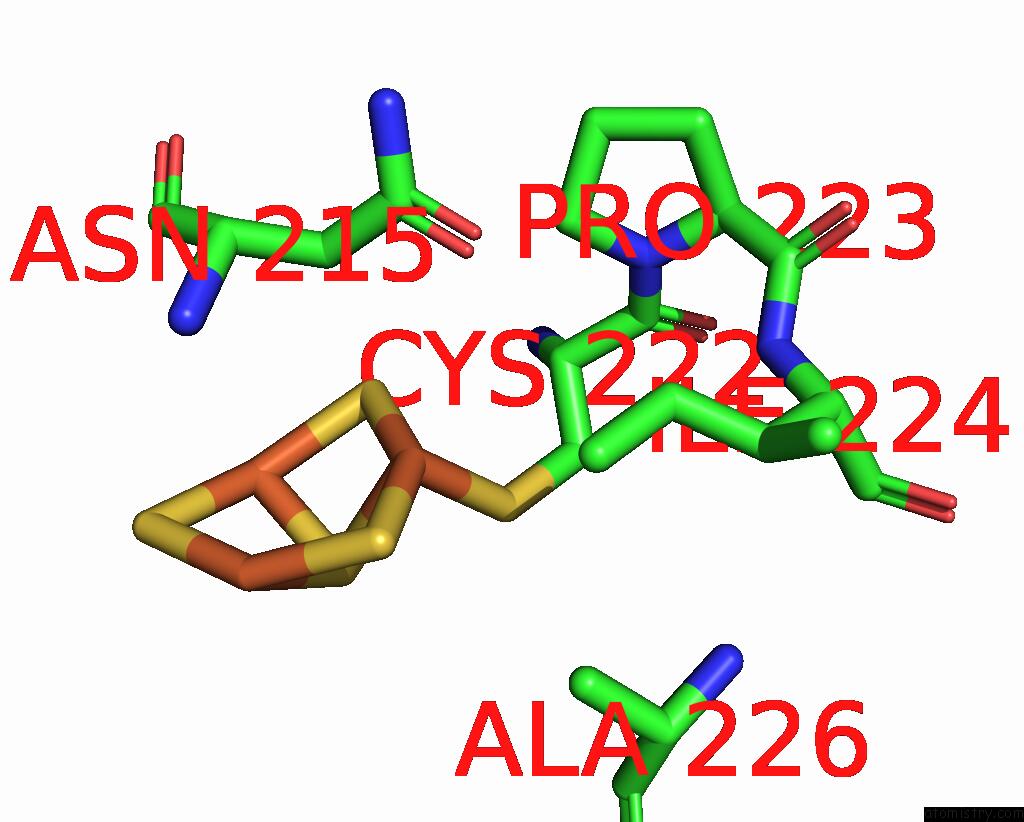
Iron binding site 2 out of 4 in 8hdd
Go back to
Iron binding site 2 out
of 4 in the Complex Structure of Catalytic, Small, and A Partial Electron Transfer Subunits From Burkholderia Cepacia Fad Glucose Dehydrogenase
Mono view

Stereo pair view
Mono view
Stereo pair view
|
|
A full contact list of Iron with other atoms in the Fe binding
site number 2 of Complex Structure of Catalytic, Small, and A Partial Electron Transfer Subunits From Burkholderia Cepacia Fad Glucose Dehydrogenase within 5.0Å range:
|
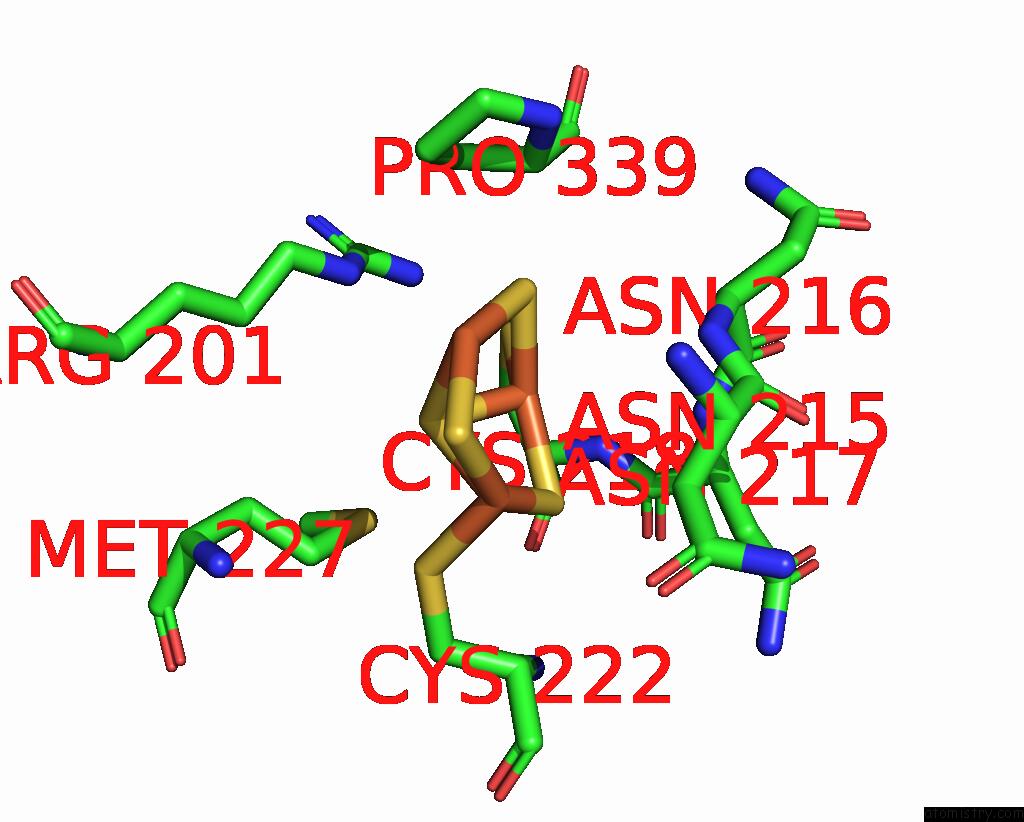
Iron binding site 3 out of 4 in 8hdd
Go back to
Iron binding site 3 out
of 4 in the Complex Structure of Catalytic, Small, and A Partial Electron Transfer Subunits From Burkholderia Cepacia Fad Glucose Dehydrogenase
Mono view

Stereo pair view
Mono view
Stereo pair view
|
|
A full contact list of Iron with other atoms in the Fe binding
site number 3 of Complex Structure of Catalytic, Small, and A Partial Electron Transfer Subunits From Burkholderia Cepacia Fad Glucose Dehydrogenase within 5.0Å range:
|
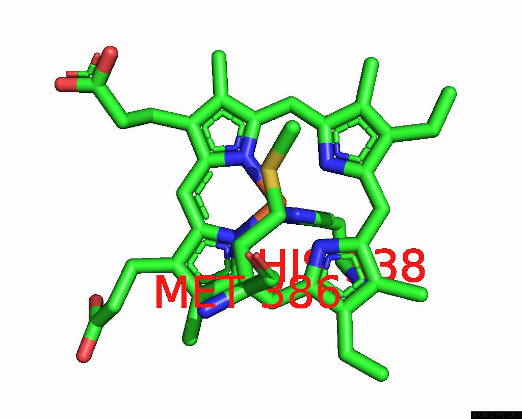
Iron binding site 4 out of 4 in 8hdd
Go back to
Iron binding site 4 out
of 4 in the Complex Structure of Catalytic, Small, and A Partial Electron Transfer Subunits From Burkholderia Cepacia Fad Glucose Dehydrogenase
Mono view
Stereo pair view
Mono view
Stereo pair view
|
|
A full contact list of Iron with other atoms in the Fe binding
site number 4 of Complex Structure of Catalytic, Small, and A Partial Electron Transfer Subunits From Burkholderia Cepacia Fad Glucose Dehydrogenase within 5.0Å range:
|
Reference:
J.Okuda-Shimazaki,
H.Yoshida,
I.Lee,
K.Kojima,
N.Suzuki,
W.Tsugawa,
M.Yamada,
K.Inaka,
H.Tanaka,
K.Sode.
Microgravity Environment Grown Crystal Structure Information Based Engineering of Direct Electron Transfer Type Glucose Dehydrogenase Commun Biol V. 5 1334 2022.
ISSN: ESSN 2399-3642
DOI: 10.1038/S42003-022-04286-9
Page generated: Thu Aug 7 17:45:28 2025
ISSN: ESSN 2399-3642
DOI: 10.1038/S42003-022-04286-9
Last articles
Mg in 9FRLMg in 9FRW
Mg in 9FHL
Mg in 9FQN
Mg in 9FQ5
Mg in 9FCO
Mg in 9FDA
Mg in 9FPY
Mg in 9FP2
Mg in 9FP0