Iron »
PDB 8hcr-8ib5 »
8hi8 »
Iron in PDB 8hi8: Crystal Structure of A Holoenzyme Tglhi with Three Fe Ions For Pseudomonas Syringae Peptidyl (S) 2-Mercaptoglycine Biosynthesis
Protein crystallography data
The structure of Crystal Structure of A Holoenzyme Tglhi with Three Fe Ions For Pseudomonas Syringae Peptidyl (S) 2-Mercaptoglycine Biosynthesis, PDB code: 8hi8
was solved by
W.Cheng,
Y.H.Zheng,
X.L.Fu,
with X-Ray Crystallography technique. A brief refinement statistics is given in the table below:
| Resolution Low / High (Å) | 29.74 / 3.49 |
| Space group | P 61 |
| Cell size a, b, c (Å), α, β, γ (°) | 118.943, 118.943, 87.46, 90, 90, 120 |
| R / Rfree (%) | 20.4 / 26 |
Iron Binding Sites:
The binding sites of Iron atom in the Crystal Structure of A Holoenzyme Tglhi with Three Fe Ions For Pseudomonas Syringae Peptidyl (S) 2-Mercaptoglycine Biosynthesis
(pdb code 8hi8). This binding sites where shown within
5.0 Angstroms radius around Iron atom.
In total 3 binding sites of Iron where determined in the Crystal Structure of A Holoenzyme Tglhi with Three Fe Ions For Pseudomonas Syringae Peptidyl (S) 2-Mercaptoglycine Biosynthesis, PDB code: 8hi8:
Jump to Iron binding site number: 1; 2; 3;
In total 3 binding sites of Iron where determined in the Crystal Structure of A Holoenzyme Tglhi with Three Fe Ions For Pseudomonas Syringae Peptidyl (S) 2-Mercaptoglycine Biosynthesis, PDB code: 8hi8:
Jump to Iron binding site number: 1; 2; 3;
Iron binding site 1 out of 3 in 8hi8
Go back to
Iron binding site 1 out
of 3 in the Crystal Structure of A Holoenzyme Tglhi with Three Fe Ions For Pseudomonas Syringae Peptidyl (S) 2-Mercaptoglycine Biosynthesis
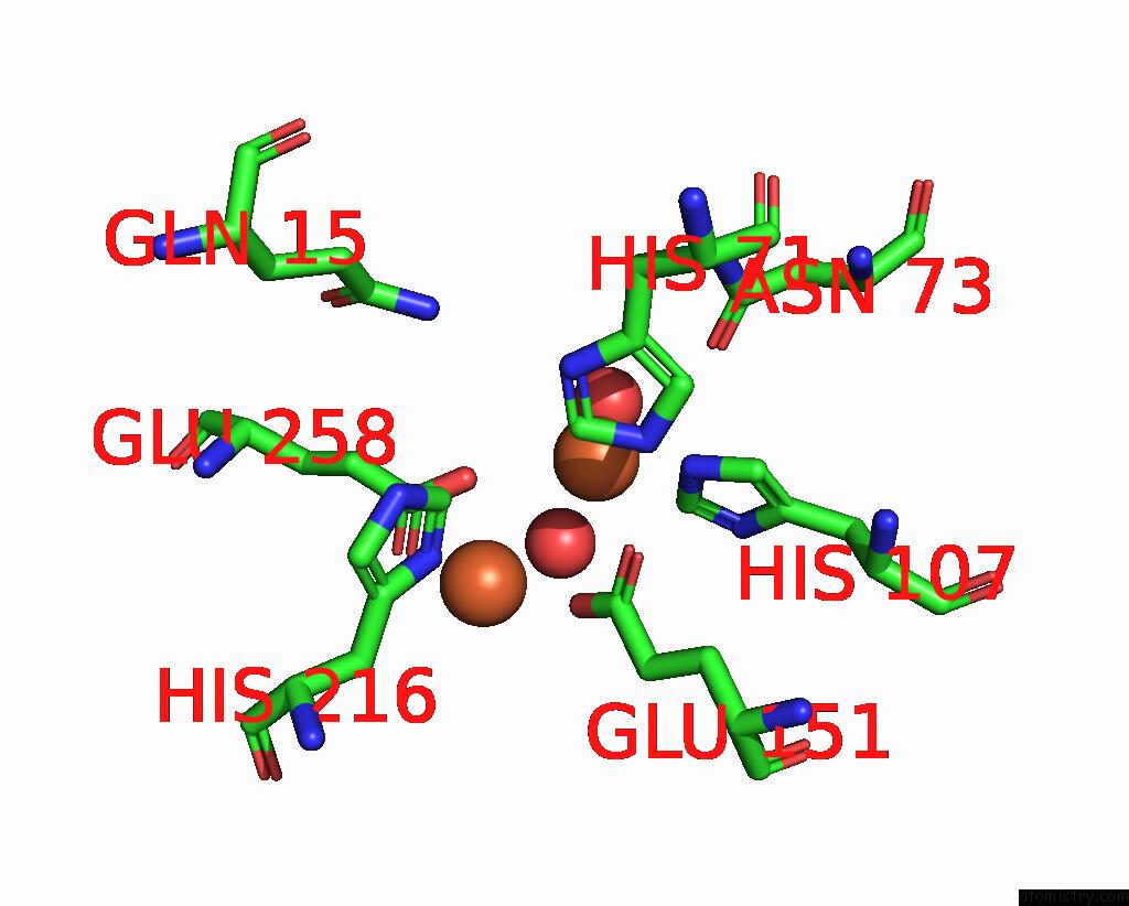
Mono view
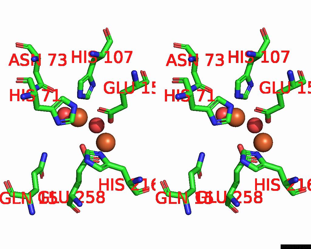
Stereo pair view
Mono view
Stereo pair view
A full contact list of Iron with other atoms in the Fe binding
site number 1 of Crystal Structure of A Holoenzyme Tglhi with Three Fe Ions For Pseudomonas Syringae Peptidyl (S) 2-Mercaptoglycine Biosynthesis within 5.0Å range:
|
Iron binding site 2 out of 3 in 8hi8
Go back to
Iron binding site 2 out
of 3 in the Crystal Structure of A Holoenzyme Tglhi with Three Fe Ions For Pseudomonas Syringae Peptidyl (S) 2-Mercaptoglycine Biosynthesis
Mono view

Stereo pair view
Mono view
Stereo pair view
A full contact list of Iron with other atoms in the Fe binding
site number 2 of Crystal Structure of A Holoenzyme Tglhi with Three Fe Ions For Pseudomonas Syringae Peptidyl (S) 2-Mercaptoglycine Biosynthesis within 5.0Å range:
|
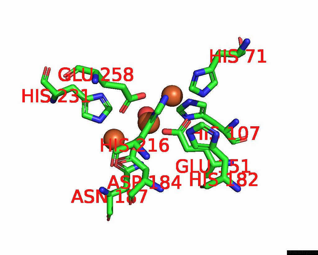
Iron binding site 3 out of 3 in 8hi8
Go back to
Iron binding site 3 out
of 3 in the Crystal Structure of A Holoenzyme Tglhi with Three Fe Ions For Pseudomonas Syringae Peptidyl (S) 2-Mercaptoglycine Biosynthesis

Mono view

Stereo pair view
Mono view
Stereo pair view
A full contact list of Iron with other atoms in the Fe binding
site number 3 of Crystal Structure of A Holoenzyme Tglhi with Three Fe Ions For Pseudomonas Syringae Peptidyl (S) 2-Mercaptoglycine Biosynthesis within 5.0Å range:
|
Reference:
W.Cheng,
Y.H.Zheng,
X.L.Fu.
Crystal Structure of A Holoenzyme Tglhi with Two Fe Irons For Pseudomonas Syringae Peptidyl (S) 2-Mercaptoglycine Biosynthesis To Be Published.
Page generated: Thu Aug 7 17:46:47 2025
Last articles
V in 1QVIV in 1QYL
V in 1NOP
V in 1MU9
V in 1LKX
V in 1M7G
V in 1M5O
V in 1DKT
V in 1JH7
V in 1J9L