Iron »
PDB 8hcr-8ib5 »
8hoq »
Iron in PDB 8hoq: Crystal Structure of the P450 BM3 Heme Domain Mutant F87A in Complex with Im-C6-Phe(4CF3)-Tyr
Enzymatic activity of Crystal Structure of the P450 BM3 Heme Domain Mutant F87A in Complex with Im-C6-Phe(4CF3)-Tyr
All present enzymatic activity of Crystal Structure of the P450 BM3 Heme Domain Mutant F87A in Complex with Im-C6-Phe(4CF3)-Tyr:
1.14.14.1; 1.6.2.4;
1.14.14.1; 1.6.2.4;
Protein crystallography data
The structure of Crystal Structure of the P450 BM3 Heme Domain Mutant F87A in Complex with Im-C6-Phe(4CF3)-Tyr, PDB code: 8hoq
was solved by
Y.Jiang,
S.Dong,
Y.Feng,
Z.Cong,
with X-Ray Crystallography technique. A brief refinement statistics is given in the table below:
| Resolution Low / High (Å) | 57.72 / 1.94 |
| Space group | P 1 21 1 |
| Cell size a, b, c (Å), α, β, γ (°) | 58.639, 147.949, 64.725, 90, 100.16, 90 |
| R / Rfree (%) | 17.8 / 20 |
Other elements in 8hoq:
The structure of Crystal Structure of the P450 BM3 Heme Domain Mutant F87A in Complex with Im-C6-Phe(4CF3)-Tyr also contains other interesting chemical elements:
| Fluorine | (F) | 6 atoms |
Iron Binding Sites:
The binding sites of Iron atom in the Crystal Structure of the P450 BM3 Heme Domain Mutant F87A in Complex with Im-C6-Phe(4CF3)-Tyr
(pdb code 8hoq). This binding sites where shown within
5.0 Angstroms radius around Iron atom.
In total 2 binding sites of Iron where determined in the Crystal Structure of the P450 BM3 Heme Domain Mutant F87A in Complex with Im-C6-Phe(4CF3)-Tyr, PDB code: 8hoq:
Jump to Iron binding site number: 1; 2;
In total 2 binding sites of Iron where determined in the Crystal Structure of the P450 BM3 Heme Domain Mutant F87A in Complex with Im-C6-Phe(4CF3)-Tyr, PDB code: 8hoq:
Jump to Iron binding site number: 1; 2;
Iron binding site 1 out of 2 in 8hoq
Go back to
Iron binding site 1 out
of 2 in the Crystal Structure of the P450 BM3 Heme Domain Mutant F87A in Complex with Im-C6-Phe(4CF3)-Tyr
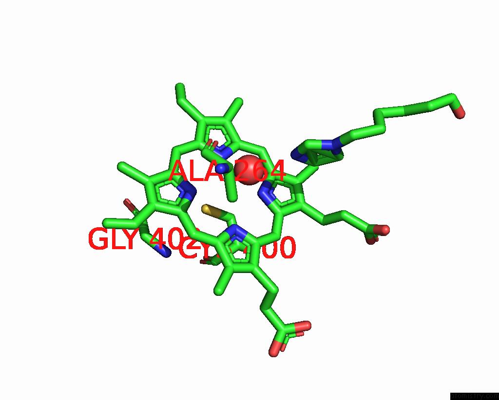
Mono view
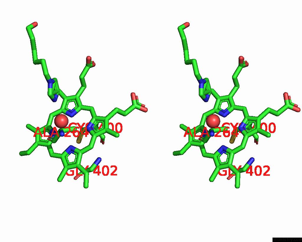
Stereo pair view
Mono view
Stereo pair view
A full contact list of Iron with other atoms in the Fe binding
site number 1 of Crystal Structure of the P450 BM3 Heme Domain Mutant F87A in Complex with Im-C6-Phe(4CF3)-Tyr within 5.0Å range:
|
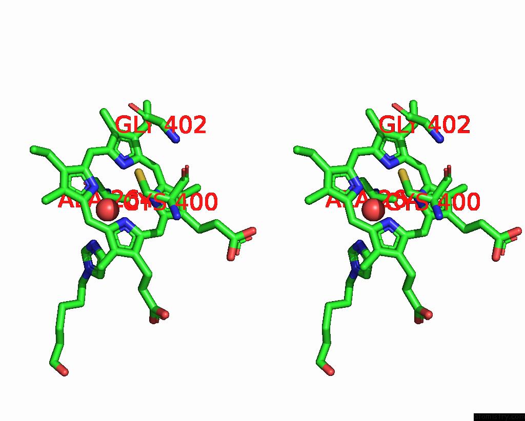
Iron binding site 2 out of 2 in 8hoq
Go back to
Iron binding site 2 out
of 2 in the Crystal Structure of the P450 BM3 Heme Domain Mutant F87A in Complex with Im-C6-Phe(4CF3)-Tyr

Mono view
Stereo pair view
Mono view
Stereo pair view
A full contact list of Iron with other atoms in the Fe binding
site number 2 of Crystal Structure of the P450 BM3 Heme Domain Mutant F87A in Complex with Im-C6-Phe(4CF3)-Tyr within 5.0Å range:
|
Reference:
Y.Jiang,
S.Dong,
Y.Feng,
Z.Cong.
Crystal Structure of the P450 BM3 Heme Domain Mutant F87A in Complex with Im-C6-Phe(4CF3)-Tyr To Be Published.
Page generated: Thu Aug 7 17:50:34 2025
Last articles
V in 1QVIV in 1QYL
V in 1NOP
V in 1MU9
V in 1LKX
V in 1M7G
V in 1M5O
V in 1DKT
V in 1JH7
V in 1J9L