Iron »
PDB 8hcr-8ib5 »
8hou »
Iron in PDB 8hou: Crystal Structure of the P450 BM3 Heme Domain Mutant F87A-T268V in Complex with Im-N-C4-Phe-Phe
Enzymatic activity of Crystal Structure of the P450 BM3 Heme Domain Mutant F87A-T268V in Complex with Im-N-C4-Phe-Phe
All present enzymatic activity of Crystal Structure of the P450 BM3 Heme Domain Mutant F87A-T268V in Complex with Im-N-C4-Phe-Phe:
1.14.14.1; 1.6.2.4;
1.14.14.1; 1.6.2.4;
Protein crystallography data
The structure of Crystal Structure of the P450 BM3 Heme Domain Mutant F87A-T268V in Complex with Im-N-C4-Phe-Phe, PDB code: 8hou
was solved by
Y.Jiang,
S.Dong,
Y.Feng,
Z.Cong,
with X-Ray Crystallography technique. A brief refinement statistics is given in the table below:
| Resolution Low / High (Å) | 57.70 / 1.75 |
| Space group | P 1 21 1 |
| Cell size a, b, c (Å), α, β, γ (°) | 58.592, 147.608, 64.764, 90, 100.03, 90 |
| R / Rfree (%) | 18.7 / 20.9 |
Iron Binding Sites:
The binding sites of Iron atom in the Crystal Structure of the P450 BM3 Heme Domain Mutant F87A-T268V in Complex with Im-N-C4-Phe-Phe
(pdb code 8hou). This binding sites where shown within
5.0 Angstroms radius around Iron atom.
In total 2 binding sites of Iron where determined in the Crystal Structure of the P450 BM3 Heme Domain Mutant F87A-T268V in Complex with Im-N-C4-Phe-Phe, PDB code: 8hou:
Jump to Iron binding site number: 1; 2;
In total 2 binding sites of Iron where determined in the Crystal Structure of the P450 BM3 Heme Domain Mutant F87A-T268V in Complex with Im-N-C4-Phe-Phe, PDB code: 8hou:
Jump to Iron binding site number: 1; 2;
Iron binding site 1 out of 2 in 8hou
Go back to
Iron binding site 1 out
of 2 in the Crystal Structure of the P450 BM3 Heme Domain Mutant F87A-T268V in Complex with Im-N-C4-Phe-Phe
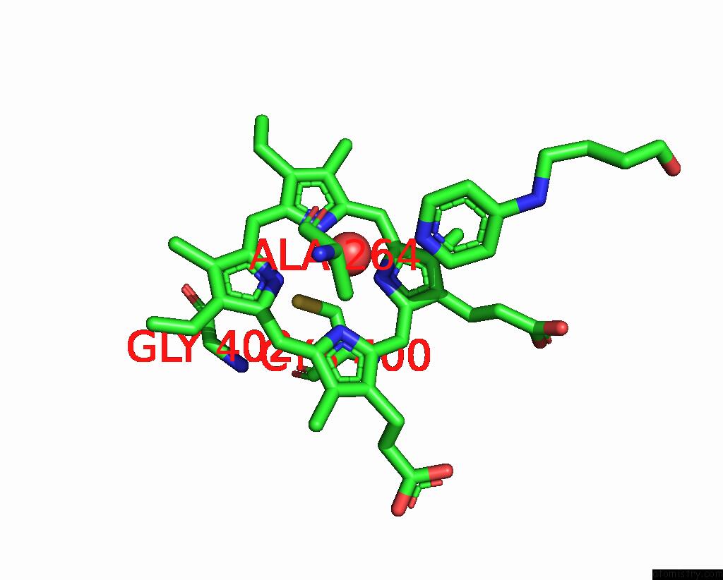
Mono view
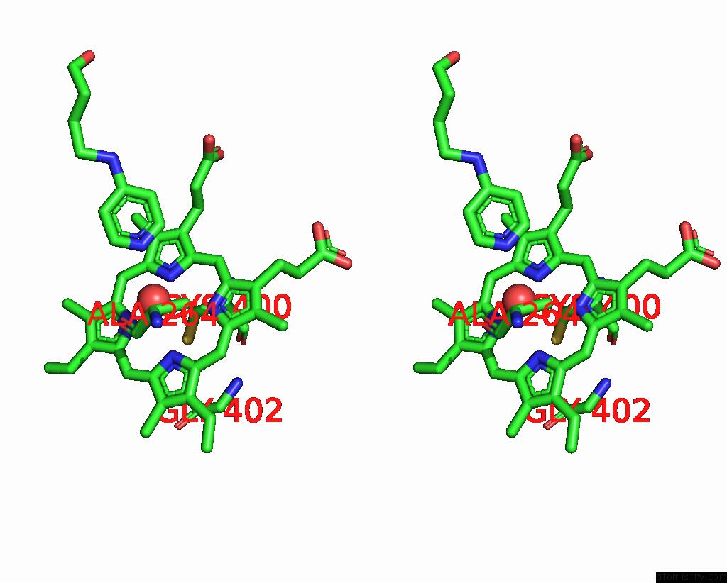
Stereo pair view
Mono view
Stereo pair view
|
|
A full contact list of Iron with other atoms in the Fe binding
site number 1 of Crystal Structure of the P450 BM3 Heme Domain Mutant F87A-T268V in Complex with Im-N-C4-Phe-Phe within 5.0Å range:
|
Iron binding site 2 out of 2 in 8hou
Go back to
Iron binding site 2 out
of 2 in the Crystal Structure of the P450 BM3 Heme Domain Mutant F87A-T268V in Complex with Im-N-C4-Phe-Phe

Mono view
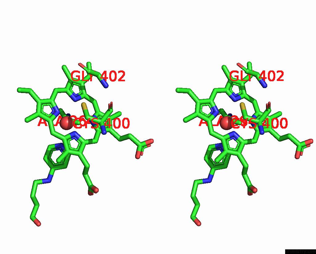
Stereo pair view
Mono view
Stereo pair view
|
|
A full contact list of Iron with other atoms in the Fe binding
site number 2 of Crystal Structure of the P450 BM3 Heme Domain Mutant F87A-T268V in Complex with Im-N-C4-Phe-Phe within 5.0Å range:
|
Reference:
Y.Jiang,
S.Dong,
Y.Feng,
Z.Cong.
Crystal Structure of the P450 BM3 Heme Domain Mutant F87A-T268V in Complex with Im-N-C4-Phe-Phe To Be Published.
Page generated: Thu Aug 7 17:51:02 2025
Last articles
Sb in 1F48Sb in 1EXI
Sb in 2W0H
Sb in 4U5T
Sb in 2XQA
Ru in 8S90
Ru in 8PH6
Ru in 8S8Z
Ru in 8RER
Ru in 8RNY