Iron »
PDB 8irg-8ji2 »
8j9l »
Iron in PDB 8j9l: Crystal Structure of Human H-Ferritin Variant 123F Assembling in SOLUTION2
Enzymatic activity of Crystal Structure of Human H-Ferritin Variant 123F Assembling in SOLUTION2
All present enzymatic activity of Crystal Structure of Human H-Ferritin Variant 123F Assembling in SOLUTION2:
1.16.3.1;
1.16.3.1;
Protein crystallography data
The structure of Crystal Structure of Human H-Ferritin Variant 123F Assembling in SOLUTION2, PDB code: 8j9l
was solved by
X.Chen,
G.Zhao,
with X-Ray Crystallography technique. A brief refinement statistics is given in the table below:
| Resolution Low / High (Å) | 29.26 / 2.50 |
| Space group | H 3 2 2 |
| Cell size a, b, c (Å), α, β, γ (°) | 261.696, 261.696, 320.511, 90, 90, 120 |
| R / Rfree (%) | 24.7 / 28.9 |
Iron Binding Sites:
Pages:
>>> Page 1 <<< Page 2, Binding sites: 11 - 16;Binding sites:
The binding sites of Iron atom in the Crystal Structure of Human H-Ferritin Variant 123F Assembling in SOLUTION2 (pdb code 8j9l). This binding sites where shown within 5.0 Angstroms radius around Iron atom.In total 16 binding sites of Iron where determined in the Crystal Structure of Human H-Ferritin Variant 123F Assembling in SOLUTION2, PDB code: 8j9l:
Jump to Iron binding site number: 1; 2; 3; 4; 5; 6; 7; 8; 9; 10;
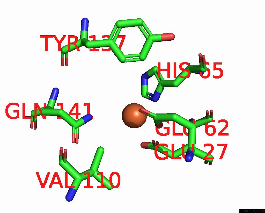
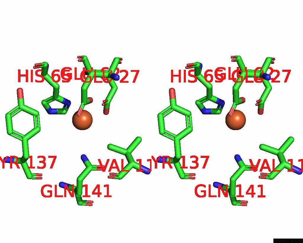
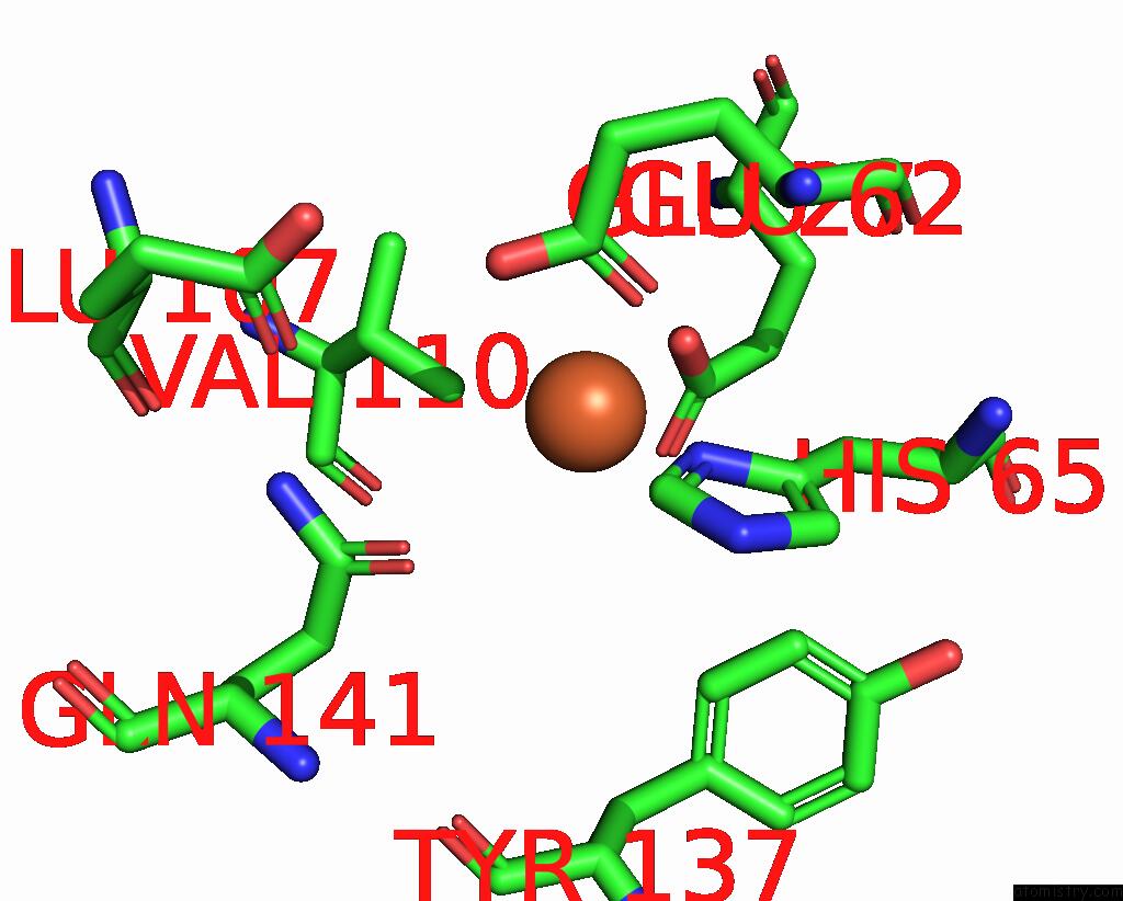
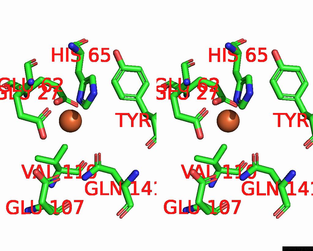
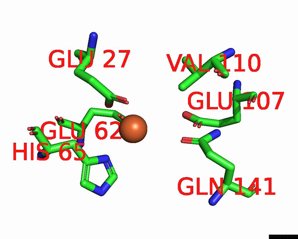
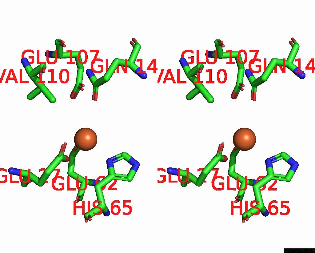
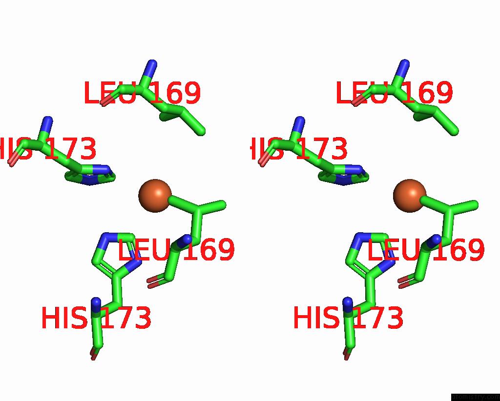
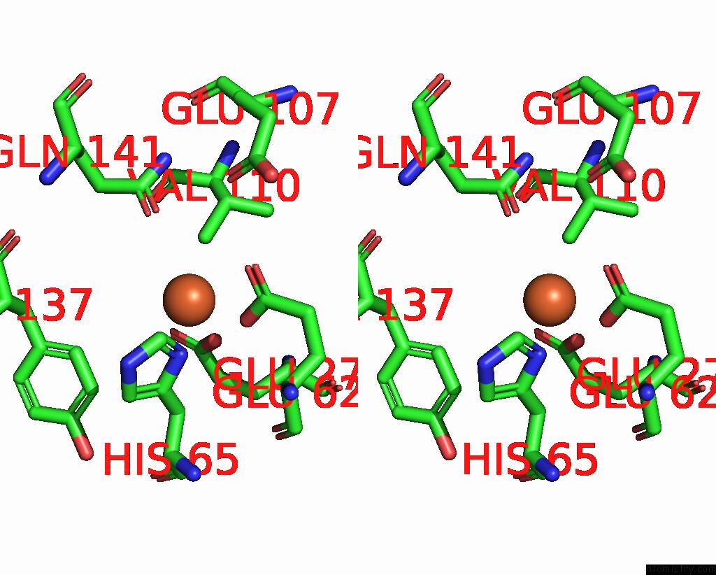
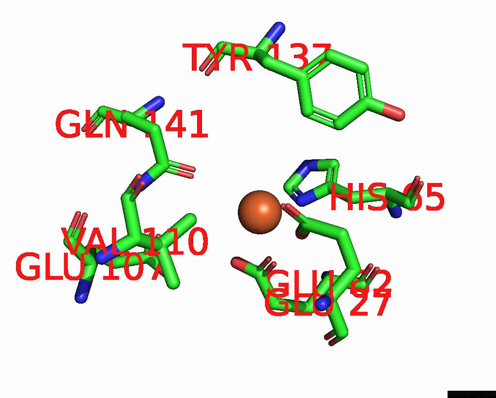
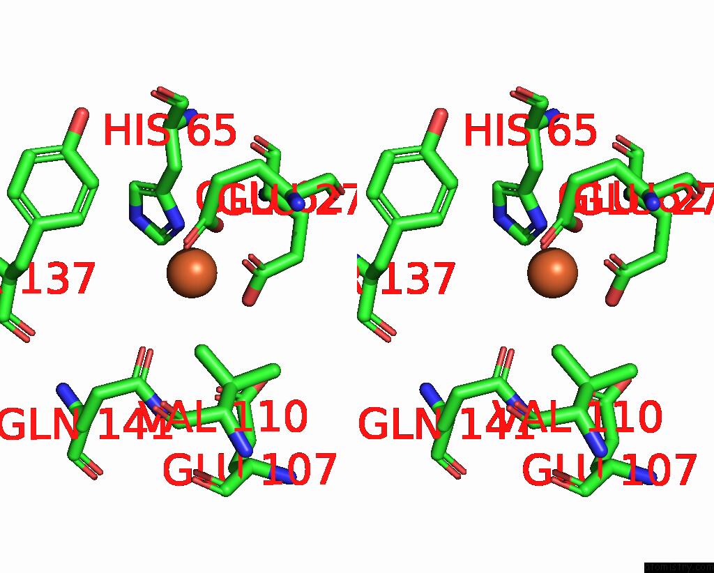
Iron binding site 1 out of 16 in 8j9l
Go back to
Iron binding site 1 out
of 16 in the Crystal Structure of Human H-Ferritin Variant 123F Assembling in SOLUTION2
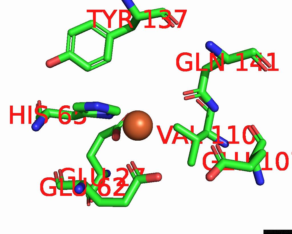
Mono view
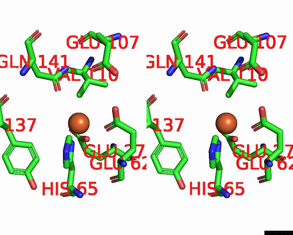
Stereo pair view
Mono view
Stereo pair view
A full contact list of Iron with other atoms in the Fe binding
site number 1 of Crystal Structure of Human H-Ferritin Variant 123F Assembling in SOLUTION2 within 5.0Å range:
|
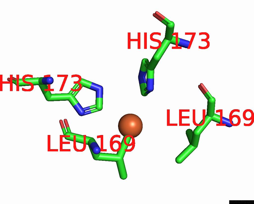
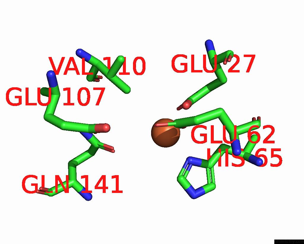
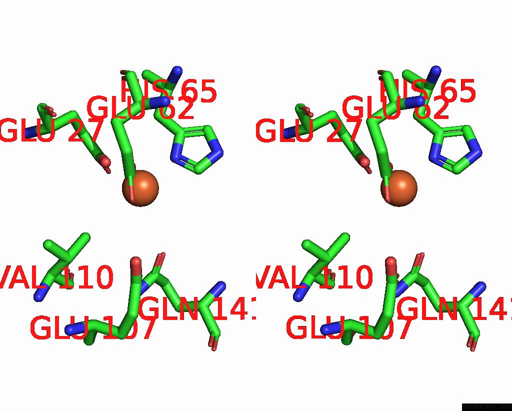
Iron binding site 2 out of 16 in 8j9l
Go back to
Iron binding site 2 out
of 16 in the Crystal Structure of Human H-Ferritin Variant 123F Assembling in SOLUTION2
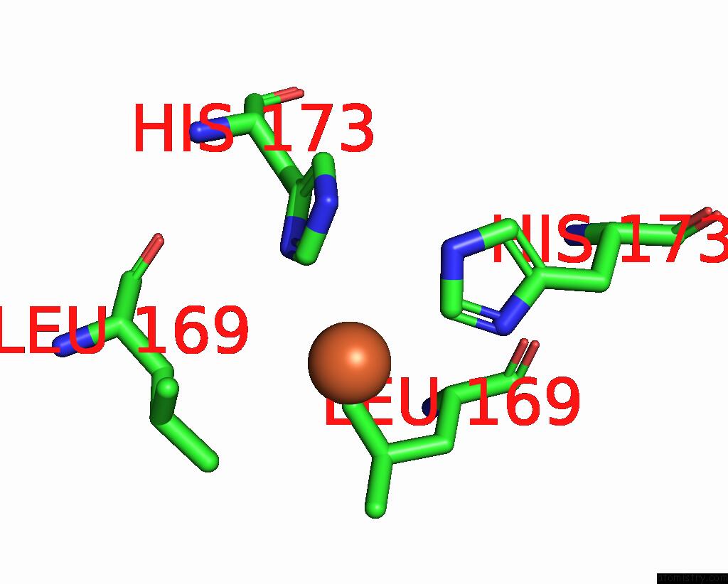
Mono view

Stereo pair view
Mono view
Stereo pair view
A full contact list of Iron with other atoms in the Fe binding
site number 2 of Crystal Structure of Human H-Ferritin Variant 123F Assembling in SOLUTION2 within 5.0Å range:
|
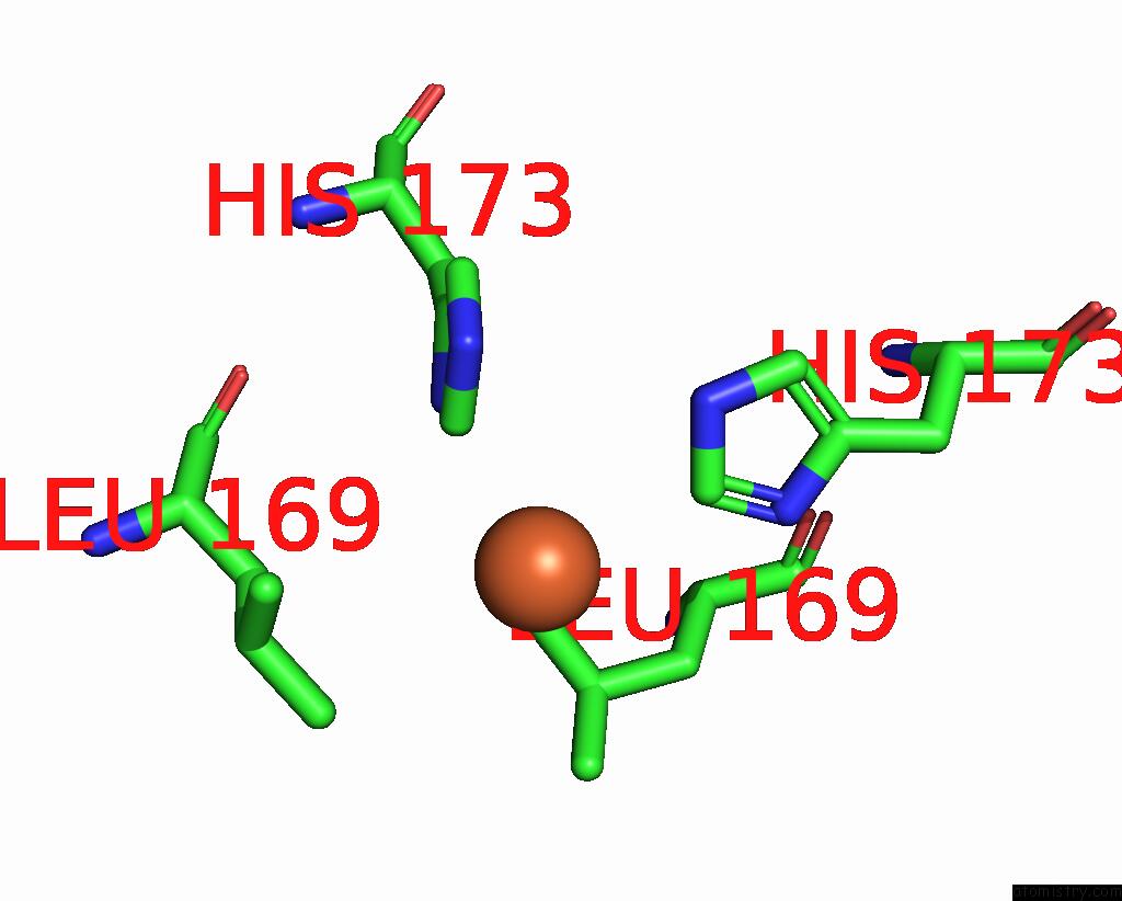
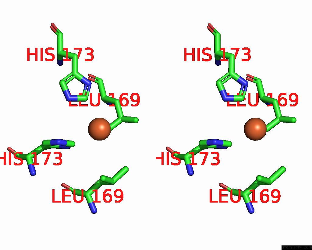
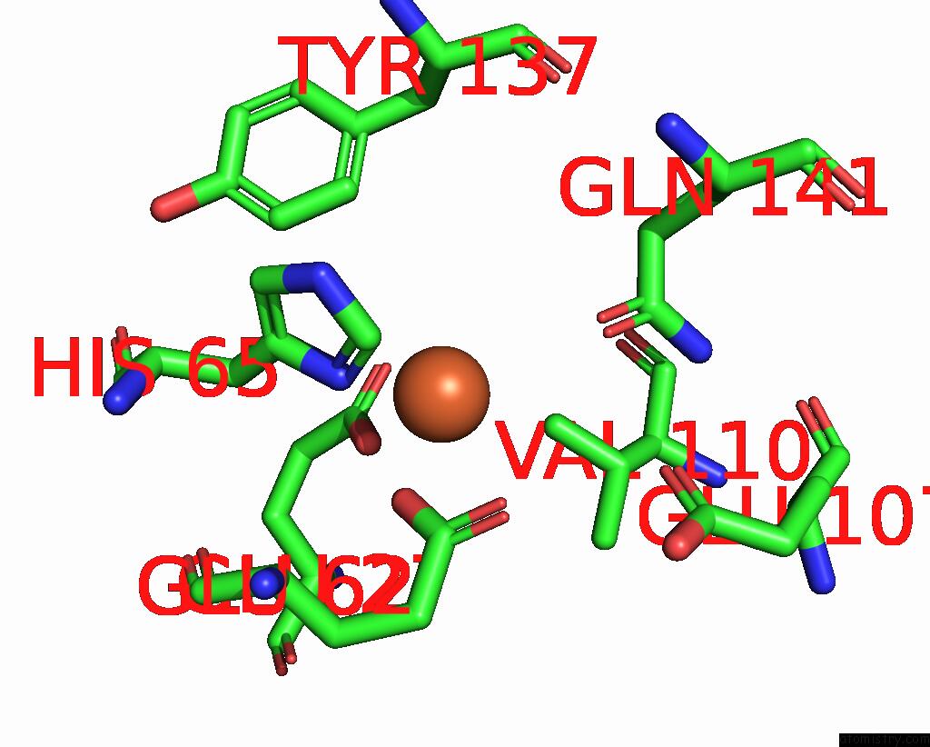
Iron binding site 3 out of 16 in 8j9l
Go back to
Iron binding site 3 out
of 16 in the Crystal Structure of Human H-Ferritin Variant 123F Assembling in SOLUTION2
Mono view
Stereo pair view
Mono view
Stereo pair view
A full contact list of Iron with other atoms in the Fe binding
site number 3 of Crystal Structure of Human H-Ferritin Variant 123F Assembling in SOLUTION2 within 5.0Å range:
|
Iron binding site 4 out of 16 in 8j9l
Go back to
Iron binding site 4 out
of 16 in the Crystal Structure of Human H-Ferritin Variant 123F Assembling in SOLUTION2
Mono view
Stereo pair view
Mono view
Stereo pair view
A full contact list of Iron with other atoms in the Fe binding
site number 4 of Crystal Structure of Human H-Ferritin Variant 123F Assembling in SOLUTION2 within 5.0Å range:
|
Iron binding site 5 out of 16 in 8j9l
Go back to
Iron binding site 5 out
of 16 in the Crystal Structure of Human H-Ferritin Variant 123F Assembling in SOLUTION2
Mono view
Stereo pair view
Mono view
Stereo pair view
A full contact list of Iron with other atoms in the Fe binding
site number 5 of Crystal Structure of Human H-Ferritin Variant 123F Assembling in SOLUTION2 within 5.0Å range:
|
Iron binding site 6 out of 16 in 8j9l
Go back to
Iron binding site 6 out
of 16 in the Crystal Structure of Human H-Ferritin Variant 123F Assembling in SOLUTION2
Mono view
Stereo pair view
Mono view
Stereo pair view
A full contact list of Iron with other atoms in the Fe binding
site number 6 of Crystal Structure of Human H-Ferritin Variant 123F Assembling in SOLUTION2 within 5.0Å range:
|
Iron binding site 7 out of 16 in 8j9l
Go back to
Iron binding site 7 out
of 16 in the Crystal Structure of Human H-Ferritin Variant 123F Assembling in SOLUTION2
Mono view
Stereo pair view
Mono view
Stereo pair view
A full contact list of Iron with other atoms in the Fe binding
site number 7 of Crystal Structure of Human H-Ferritin Variant 123F Assembling in SOLUTION2 within 5.0Å range:
|
Iron binding site 8 out of 16 in 8j9l
Go back to
Iron binding site 8 out
of 16 in the Crystal Structure of Human H-Ferritin Variant 123F Assembling in SOLUTION2
Mono view
Stereo pair view
Mono view
Stereo pair view
A full contact list of Iron with other atoms in the Fe binding
site number 8 of Crystal Structure of Human H-Ferritin Variant 123F Assembling in SOLUTION2 within 5.0Å range:
|
Iron binding site 9 out of 16 in 8j9l
Go back to
Iron binding site 9 out
of 16 in the Crystal Structure of Human H-Ferritin Variant 123F Assembling in SOLUTION2
Mono view
Stereo pair view
Mono view
Stereo pair view
A full contact list of Iron with other atoms in the Fe binding
site number 9 of Crystal Structure of Human H-Ferritin Variant 123F Assembling in SOLUTION2 within 5.0Å range:
|
Iron binding site 10 out of 16 in 8j9l
Go back to
Iron binding site 10 out
of 16 in the Crystal Structure of Human H-Ferritin Variant 123F Assembling in SOLUTION2
Mono view
Stereo pair view
Mono view
Stereo pair view
A full contact list of Iron with other atoms in the Fe binding
site number 10 of Crystal Structure of Human H-Ferritin Variant 123F Assembling in SOLUTION2 within 5.0Å range:
|
Reference:
X.Chen,
T.Zhang,
H.Liu,
J.Zang,
C.Lv,
M.Du,
G.Zhao.
Shape-Anisotropic Assembly of Protein Nanocages with Identical Building Blocks By Designed Intermolecular Pi-Pi Interactions. Adv Sci V. 10 05398 2023.
ISSN: ESSN 2198-3844
PubMed: 37870198
DOI: 10.1002/ADVS.202305398
Page generated: Thu Aug 7 18:33:43 2025
ISSN: ESSN 2198-3844
PubMed: 37870198
DOI: 10.1002/ADVS.202305398
Last articles
Mn in 9LJUMn in 9LJW
Mn in 9LJS
Mn in 9LJR
Mn in 9LJT
Mn in 9LJV
Mg in 9UA2
Mg in 9R96
Mg in 9VM1
Mg in 9P01