Iron »
PDB 8ji3-8k6k »
8joo »
Iron in PDB 8joo: Crystal Structure of Cytochrome P450 Ikad From Streptomyces Sp. ZJ306, in Complex with the Substrate Ikarugamycin
Protein crystallography data
The structure of Crystal Structure of Cytochrome P450 Ikad From Streptomyces Sp. ZJ306, in Complex with the Substrate Ikarugamycin, PDB code: 8joo
was solved by
Y.L.Zhang,
L.P.Zhang,
C.S.Zhang,
with X-Ray Crystallography technique. A brief refinement statistics is given in the table below:
| Resolution Low / High (Å) | 19.89 / 2.25 |
| Space group | I 1 2 1 |
| Cell size a, b, c (Å), α, β, γ (°) | 92.174, 82.121, 143.92, 90, 94.41, 90 |
| R / Rfree (%) | 21.7 / 26.4 |
Other elements in 8joo:
The structure of Crystal Structure of Cytochrome P450 Ikad From Streptomyces Sp. ZJ306, in Complex with the Substrate Ikarugamycin also contains other interesting chemical elements:
| Sodium | (Na) | 1 atom |
Iron Binding Sites:
The binding sites of Iron atom in the Crystal Structure of Cytochrome P450 Ikad From Streptomyces Sp. ZJ306, in Complex with the Substrate Ikarugamycin
(pdb code 8joo). This binding sites where shown within
5.0 Angstroms radius around Iron atom.
In total 2 binding sites of Iron where determined in the Crystal Structure of Cytochrome P450 Ikad From Streptomyces Sp. ZJ306, in Complex with the Substrate Ikarugamycin, PDB code: 8joo:
Jump to Iron binding site number: 1; 2;
In total 2 binding sites of Iron where determined in the Crystal Structure of Cytochrome P450 Ikad From Streptomyces Sp. ZJ306, in Complex with the Substrate Ikarugamycin, PDB code: 8joo:
Jump to Iron binding site number: 1; 2;
Iron binding site 1 out of 2 in 8joo
Go back to
Iron binding site 1 out
of 2 in the Crystal Structure of Cytochrome P450 Ikad From Streptomyces Sp. ZJ306, in Complex with the Substrate Ikarugamycin
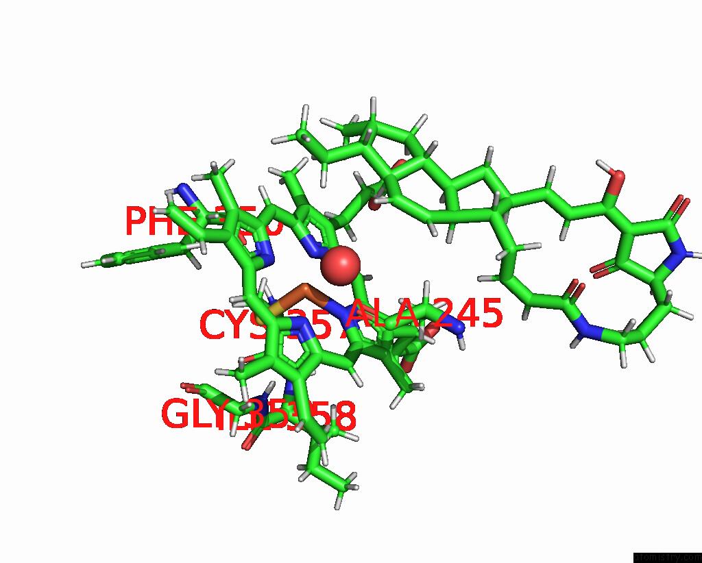
Mono view
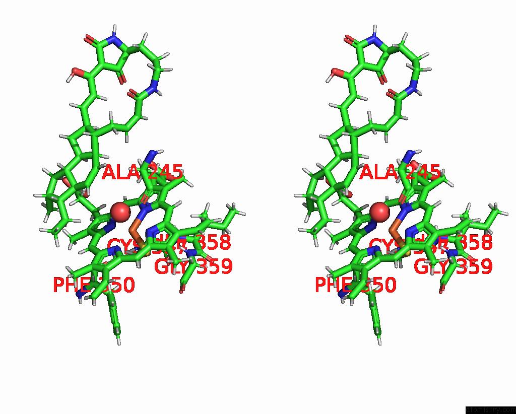
Stereo pair view
Mono view
Stereo pair view
A full contact list of Iron with other atoms in the Fe binding
site number 1 of Crystal Structure of Cytochrome P450 Ikad From Streptomyces Sp. ZJ306, in Complex with the Substrate Ikarugamycin within 5.0Å range:
|
Iron binding site 2 out of 2 in 8joo
Go back to
Iron binding site 2 out
of 2 in the Crystal Structure of Cytochrome P450 Ikad From Streptomyces Sp. ZJ306, in Complex with the Substrate Ikarugamycin
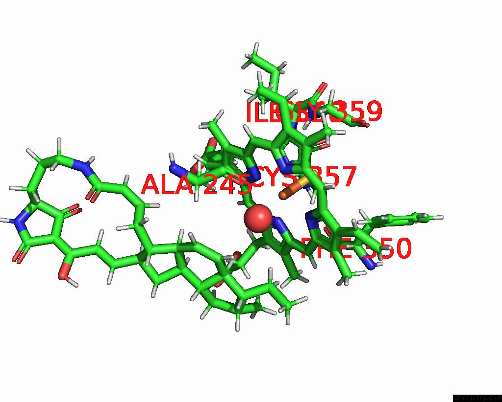
Mono view
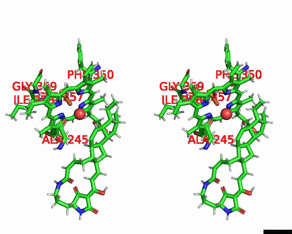
Stereo pair view
Mono view
Stereo pair view
A full contact list of Iron with other atoms in the Fe binding
site number 2 of Crystal Structure of Cytochrome P450 Ikad From Streptomyces Sp. ZJ306, in Complex with the Substrate Ikarugamycin within 5.0Å range:
|
Reference:
P.Jiang,
H.Jin,
G.Zhang,
W.Zhang,
W.Liu,
Y.Zhu,
C.Zhang,
L.Zhang.
A Mechanistic Understanding of the Distinct Regio- and Chemoselectivity of Multifunctional P450S By Structural Comparison of Ikad and Cfta Complexed with Common Substrates. Angew.Chem.Int.Ed.Engl. 10728 2023.
ISSN: ESSN 1521-3773
PubMed: 37917570
DOI: 10.1002/ANIE.202310728
Page generated: Thu Aug 7 18:47:03 2025
ISSN: ESSN 1521-3773
PubMed: 37917570
DOI: 10.1002/ANIE.202310728
Last articles
Mn in 9LJUMn in 9LJW
Mn in 9LJS
Mn in 9LJR
Mn in 9LJT
Mn in 9LJV
Mg in 9UA2
Mg in 9R96
Mg in 9VM1
Mg in 9P01