Iron »
PDB 8k6y-8p02 »
8ogg »
Iron in PDB 8ogg: Crystal Structure of Futa After An Accumulated Dose of 5 Kgy
Protein crystallography data
The structure of Crystal Structure of Futa After An Accumulated Dose of 5 Kgy, PDB code: 8ogg
was solved by
R.Bolton,
I.Tews,
with X-Ray Crystallography technique. A brief refinement statistics is given in the table below:
| Resolution Low / High (Å) | 40.97 / 1.76 |
| Space group | P 1 21 1 |
| Cell size a, b, c (Å), α, β, γ (°) | 39.718, 78.66, 48.382, 90, 97.79, 90 |
| R / Rfree (%) | 20.2 / 24.1 |
Iron Binding Sites:
The binding sites of Iron atom in the Crystal Structure of Futa After An Accumulated Dose of 5 Kgy
(pdb code 8ogg). This binding sites where shown within
5.0 Angstroms radius around Iron atom.
In total only one binding site of Iron was determined in the Crystal Structure of Futa After An Accumulated Dose of 5 Kgy, PDB code: 8ogg:
In total only one binding site of Iron was determined in the Crystal Structure of Futa After An Accumulated Dose of 5 Kgy, PDB code: 8ogg:
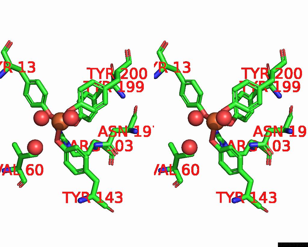
Iron binding site 1 out of 1 in 8ogg
Go back to
Iron binding site 1 out
of 1 in the Crystal Structure of Futa After An Accumulated Dose of 5 Kgy

Mono view
Stereo pair view
Mono view
Stereo pair view
A full contact list of Iron with other atoms in the Fe binding
site number 1 of Crystal Structure of Futa After An Accumulated Dose of 5 Kgy within 5.0Å range:
|
Reference:
R.Bolton,
I.Tews.
A Redox Switch Allows Binding of Ferrous and Ferric Ions in the Cyanobacterial Iron Binding Protein Futa From Prochlorococcus Biorxiv 2023.
ISSN: ISSN 2692-8205
DOI: 10.1101/2023.05.23.541926V1
Page generated: Thu Aug 7 18:58:32 2025
ISSN: ISSN 2692-8205
DOI: 10.1101/2023.05.23.541926V1
Last articles
Mg in 5YEUMg in 5YFM
Mg in 5YEW
Mg in 5YEQ
Mg in 5Y5S
Mg in 5YEC
Mg in 5YEE
Mg in 5Y9E
Mg in 5YD6
Mg in 5YCD