Iron »
PDB 8k6y-8p02 »
8ogi »
Iron in PDB 8ogi: Structure of Native Human Eosinophil Peroxidase
Protein crystallography data
The structure of Structure of Native Human Eosinophil Peroxidase, PDB code: 8ogi
was solved by
V.Pfanzagl,
C.Obinger,
with X-Ray Crystallography technique. A brief refinement statistics is given in the table below:
| Resolution Low / High (Å) | 22.57 / 1.55 |
| Space group | P 21 21 21 |
| Cell size a, b, c (Å), α, β, γ (°) | 53.123, 85.562, 139.395, 90, 90, 90 |
| R / Rfree (%) | 17.4 / 18.5 |
Other elements in 8ogi:
The structure of Structure of Native Human Eosinophil Peroxidase also contains other interesting chemical elements:
| Chlorine | (Cl) | 2 atoms |
| Calcium | (Ca) | 1 atom |
Iron Binding Sites:
The binding sites of Iron atom in the Structure of Native Human Eosinophil Peroxidase
(pdb code 8ogi). This binding sites where shown within
5.0 Angstroms radius around Iron atom.
In total only one binding site of Iron was determined in the Structure of Native Human Eosinophil Peroxidase, PDB code: 8ogi:
In total only one binding site of Iron was determined in the Structure of Native Human Eosinophil Peroxidase, PDB code: 8ogi:
Iron binding site 1 out of 1 in 8ogi
Go back to
Iron binding site 1 out
of 1 in the Structure of Native Human Eosinophil Peroxidase
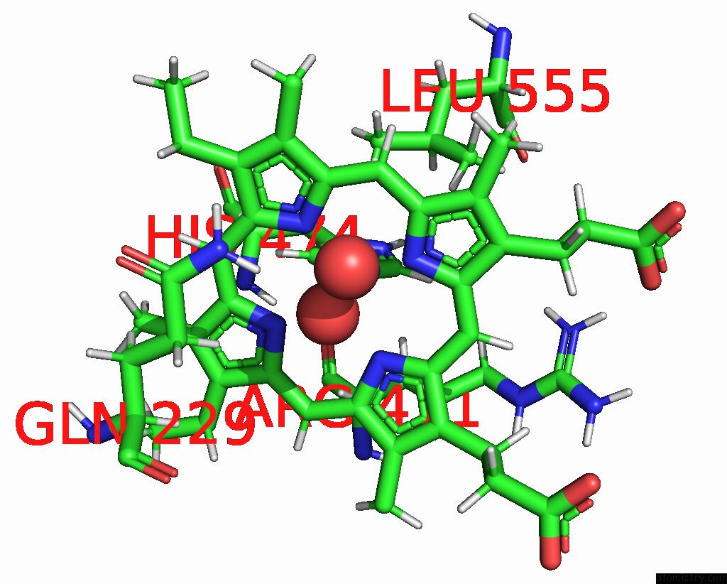
Mono view
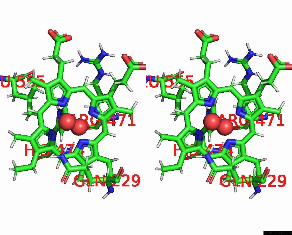
Stereo pair view
Mono view
Stereo pair view
|
|
A full contact list of Iron with other atoms in the Fe binding
site number 1 of Structure of Native Human Eosinophil Peroxidase within 5.0Å range:
|
Reference:
V.Pfanzagl,
C.Gruber-Grunwald,
U.Leitgeb,
P.G.Furtmuller,
C.Obinger.
Posttranslational Modification and Heme Cavity Architecture of Human Eosinophil Peroxidase-Insights From First Crystal Structure and Biochemical Characterization. J.Biol.Chem. V. 299 05402 2023.
ISSN: ESSN 1083-351X
PubMed: 38229400
DOI: 10.1016/J.JBC.2023.105402
Page generated: Thu Aug 7 18:58:50 2025
ISSN: ESSN 1083-351X
PubMed: 38229400
DOI: 10.1016/J.JBC.2023.105402
Last articles
Zn in 2XX7Zn in 2XX0
Zn in 2XWR
Zn in 2XVA
Zn in 2XWC
Zn in 2XSN
Zn in 2XUE
Zn in 2XUM
Zn in 2XS3
Zn in 2XSC