Iron »
PDB 8rgt-8rxn »
8rtv »
Iron in PDB 8rtv: Crystal Structure of CYP154E1 From Thermobifida Fusca Yx in Complex with 4-Phenylimidazole
Protein crystallography data
The structure of Crystal Structure of CYP154E1 From Thermobifida Fusca Yx in Complex with 4-Phenylimidazole, PDB code: 8rtv
was solved by
K.Bikbaev,
S.Hoelzel,
V.Urlacher,
I.Span,
with X-Ray Crystallography technique. A brief refinement statistics is given in the table below:
| Resolution Low / High (Å) | 48.98 / 2.00 |
| Space group | P 21 21 21 |
| Cell size a, b, c (Å), α, β, γ (°) | 62.334, 97.176, 135.688, 90, 90, 90 |
| R / Rfree (%) | 21.8 / 27.1 |
Iron Binding Sites:
The binding sites of Iron atom in the Crystal Structure of CYP154E1 From Thermobifida Fusca Yx in Complex with 4-Phenylimidazole
(pdb code 8rtv). This binding sites where shown within
5.0 Angstroms radius around Iron atom.
In total 2 binding sites of Iron where determined in the Crystal Structure of CYP154E1 From Thermobifida Fusca Yx in Complex with 4-Phenylimidazole, PDB code: 8rtv:
Jump to Iron binding site number: 1; 2;
In total 2 binding sites of Iron where determined in the Crystal Structure of CYP154E1 From Thermobifida Fusca Yx in Complex with 4-Phenylimidazole, PDB code: 8rtv:
Jump to Iron binding site number: 1; 2;
Iron binding site 1 out of 2 in 8rtv
Go back to
Iron binding site 1 out
of 2 in the Crystal Structure of CYP154E1 From Thermobifida Fusca Yx in Complex with 4-Phenylimidazole
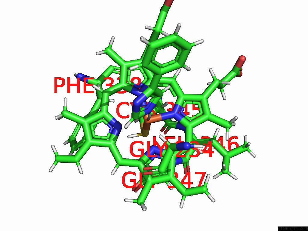
Mono view
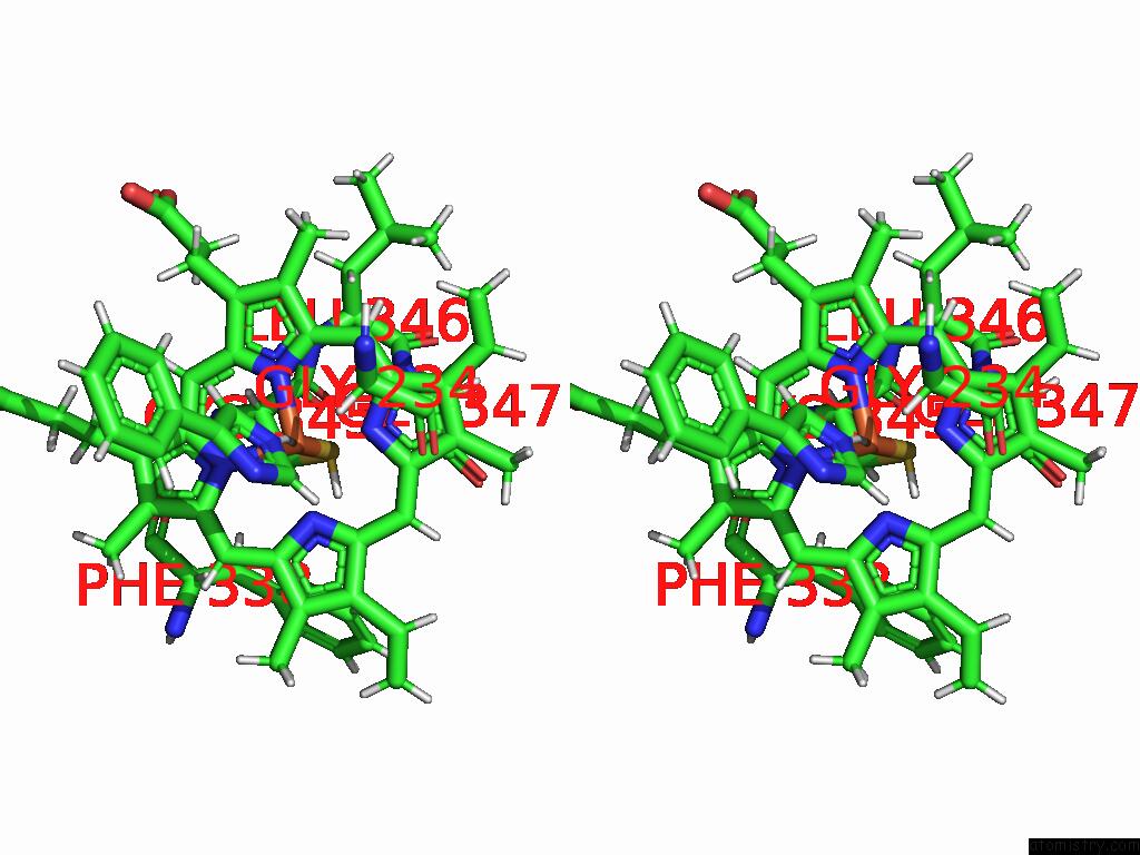
Stereo pair view
Mono view
Stereo pair view
A full contact list of Iron with other atoms in the Fe binding
site number 1 of Crystal Structure of CYP154E1 From Thermobifida Fusca Yx in Complex with 4-Phenylimidazole within 5.0Å range:
|
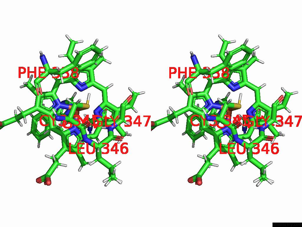
Iron binding site 2 out of 2 in 8rtv
Go back to
Iron binding site 2 out
of 2 in the Crystal Structure of CYP154E1 From Thermobifida Fusca Yx in Complex with 4-Phenylimidazole
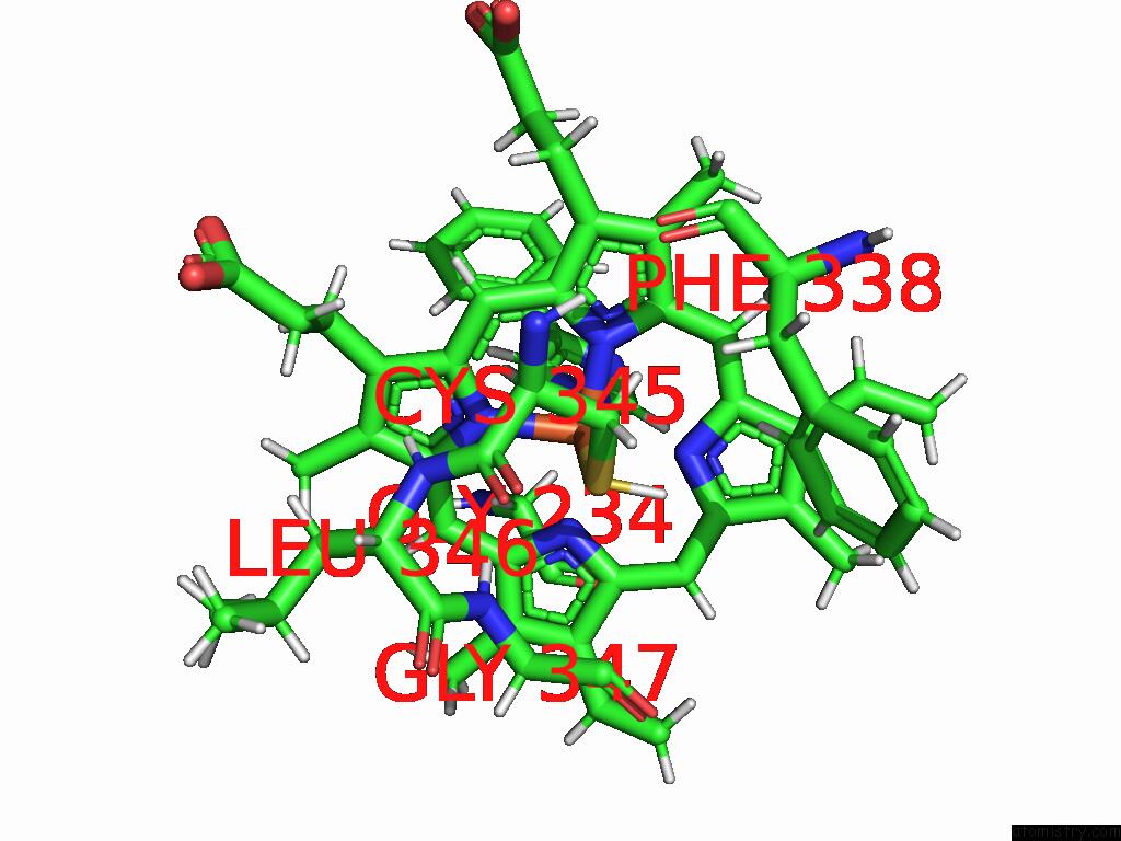
Mono view
Stereo pair view
Mono view
Stereo pair view
A full contact list of Iron with other atoms in the Fe binding
site number 2 of Crystal Structure of CYP154E1 From Thermobifida Fusca Yx in Complex with 4-Phenylimidazole within 5.0Å range:
|
Reference:
K.Bikbaev,
S.Hoelzel,
V.Urlacher,
I.Span.
Structural Determination of the CYP154E1 Cytochrome P450 From Thermobifida Fusca Yx To Be Published.
Page generated: Thu Aug 7 22:33:53 2025
Last articles
V in 1RXSV in 1UZI
V in 1YV3
V in 1VOM
V in 1VNI
V in 1VNH
V in 1VNG
V in 1VNF
V in 1VNE
V in 1VNC