Iron »
PDB 8rgt-8s5h »
8s53 »
Iron in PDB 8s53: X-Ray Crystal Structure of CYP142 From Mycobacterium Tuberculosis in Complex with A Fragment Bound in Two Poses
Enzymatic activity of X-Ray Crystal Structure of CYP142 From Mycobacterium Tuberculosis in Complex with A Fragment Bound in Two Poses
All present enzymatic activity of X-Ray Crystal Structure of CYP142 From Mycobacterium Tuberculosis in Complex with A Fragment Bound in Two Poses:
1.14.15.28;
1.14.15.28;
Protein crystallography data
The structure of X-Ray Crystal Structure of CYP142 From Mycobacterium Tuberculosis in Complex with A Fragment Bound in Two Poses, PDB code: 8s53
was solved by
M.Snee,
M.Kavanagh,
C.Levy,
D.Leys,
with X-Ray Crystallography technique. A brief refinement statistics is given in the table below:
| Resolution Low / High (Å) | 65.33 / 1.60 |
| Space group | P 21 21 21 |
| Cell size a, b, c (Å), α, β, γ (°) | 55.458, 65.772, 130.655, 90, 90, 90 |
| R / Rfree (%) | 15.6 / 17.5 |
Iron Binding Sites:
The binding sites of Iron atom in the X-Ray Crystal Structure of CYP142 From Mycobacterium Tuberculosis in Complex with A Fragment Bound in Two Poses
(pdb code 8s53). This binding sites where shown within
5.0 Angstroms radius around Iron atom.
In total only one binding site of Iron was determined in the X-Ray Crystal Structure of CYP142 From Mycobacterium Tuberculosis in Complex with A Fragment Bound in Two Poses, PDB code: 8s53:
In total only one binding site of Iron was determined in the X-Ray Crystal Structure of CYP142 From Mycobacterium Tuberculosis in Complex with A Fragment Bound in Two Poses, PDB code: 8s53:
Iron binding site 1 out of 1 in 8s53
Go back to
Iron binding site 1 out
of 1 in the X-Ray Crystal Structure of CYP142 From Mycobacterium Tuberculosis in Complex with A Fragment Bound in Two Poses
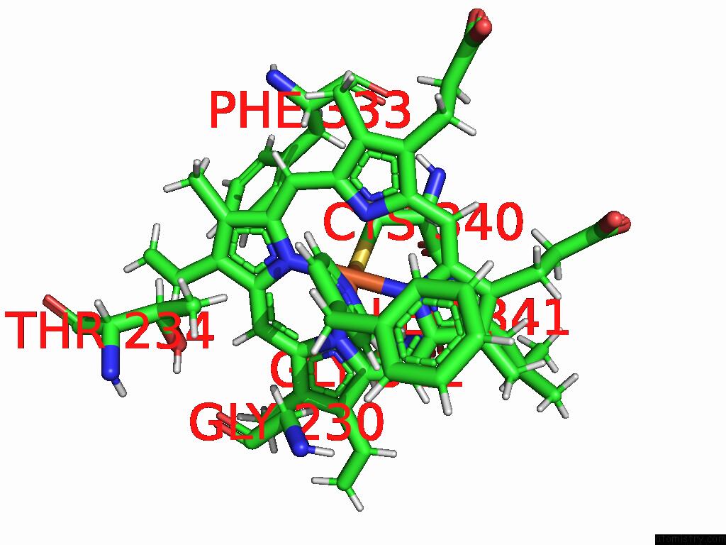
Mono view
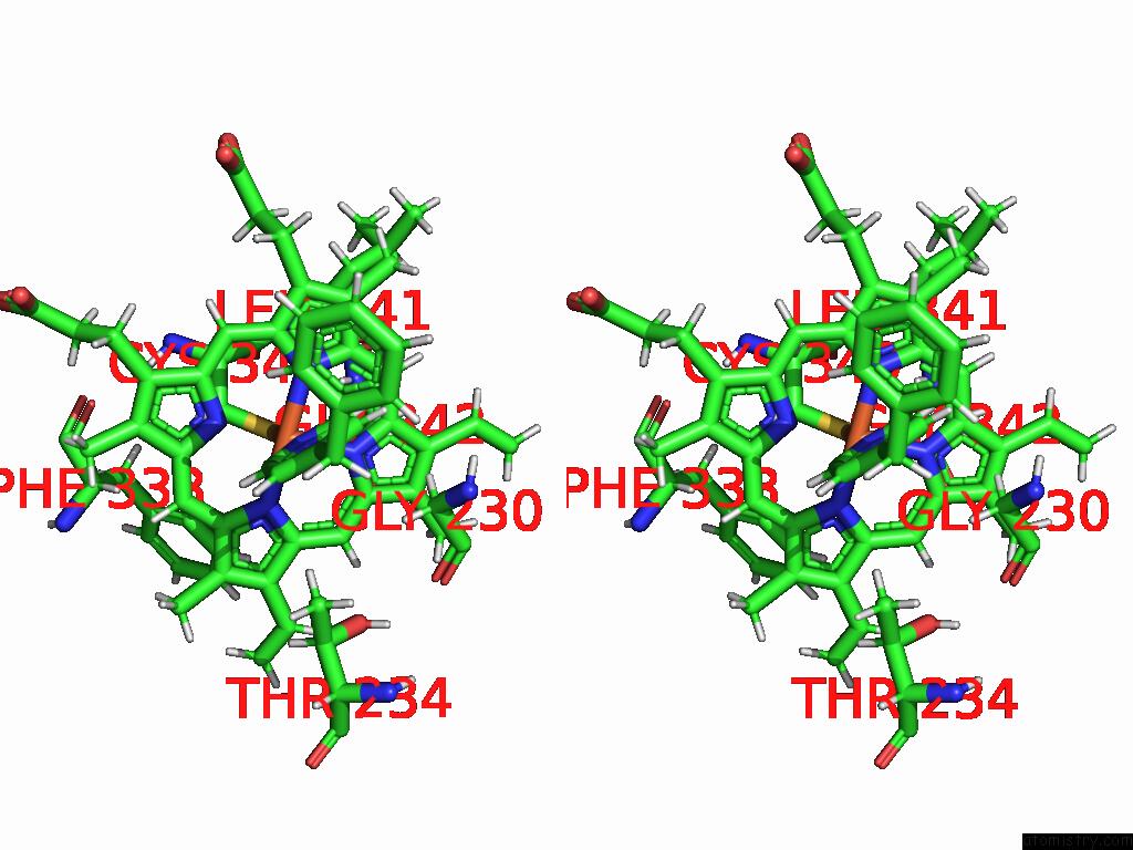
Stereo pair view
Mono view
Stereo pair view
A full contact list of Iron with other atoms in the Fe binding
site number 1 of X-Ray Crystal Structure of CYP142 From Mycobacterium Tuberculosis in Complex with A Fragment Bound in Two Poses within 5.0Å range:
|
Reference:
M.E.Kavanagh,
K.J.Mclean,
S.H.Gilbert,
C.Amadi,
M.Snee,
R.B.Tunnicliffe,
K.Arora,
H.I.Boshoff,
A.Fanourakis,
M.J.Rebello-Lopez,
F.Ortega-Muro,
C.W.Levy,
A.W.Munro,
D.Leys,
C.Abell,
A.G.Coyne.
Fragment-Based Development of Small Molecule Inhibitors Targeting Mycobacterium Tuberculosis Cholesterol Metabolism. Biorxiv 2024.
ISSN: ISSN 2692-8205
PubMed: 39803573
DOI: 10.1101/2024.10.28.620643
Page generated: Sun Feb 9 07:20:48 2025
ISSN: ISSN 2692-8205
PubMed: 39803573
DOI: 10.1101/2024.10.28.620643
Last articles
Fe in 2YXOFe in 2YRS
Fe in 2YXC
Fe in 2YNM
Fe in 2YVJ
Fe in 2YP1
Fe in 2YU2
Fe in 2YU1
Fe in 2YQB
Fe in 2YOO