Iron »
PDB 8u1i-8uoq »
8ugo »
Iron in PDB 8ugo: Structure of the Complex Between Human Lias and H-Protein in the Presence of 5'-Deoxyadenosine
Enzymatic activity of Structure of the Complex Between Human Lias and H-Protein in the Presence of 5'-Deoxyadenosine
All present enzymatic activity of Structure of the Complex Between Human Lias and H-Protein in the Presence of 5'-Deoxyadenosine:
2.8.1.8;
2.8.1.8;
Protein crystallography data
The structure of Structure of the Complex Between Human Lias and H-Protein in the Presence of 5'-Deoxyadenosine, PDB code: 8ugo
was solved by
O.A.Esakova,
D.M.Warui,
S.S.Neti,
J.N.Alumasa,
S.J.Booker,
with X-Ray Crystallography technique. A brief refinement statistics is given in the table below:
| Resolution Low / High (Å) | 38.06 / 2.45 |
| Space group | P 21 21 21 |
| Cell size a, b, c (Å), α, β, γ (°) | 48.681, 169.209, 183.183, 90, 90, 90 |
| R / Rfree (%) | 18.5 / 22.3 |
Iron Binding Sites:
Pages:
>>> Page 1 <<< Page 2, Binding sites: 11 - 20; Page 3, Binding sites: 21 - 21;Binding sites:
The binding sites of Iron atom in the Structure of the Complex Between Human Lias and H-Protein in the Presence of 5'-Deoxyadenosine (pdb code 8ugo). This binding sites where shown within 5.0 Angstroms radius around Iron atom.In total 21 binding sites of Iron where determined in the Structure of the Complex Between Human Lias and H-Protein in the Presence of 5'-Deoxyadenosine, PDB code: 8ugo:
Jump to Iron binding site number: 1; 2; 3; 4; 5; 6; 7; 8; 9; 10;
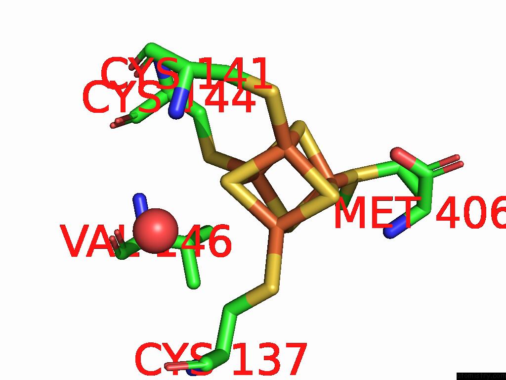
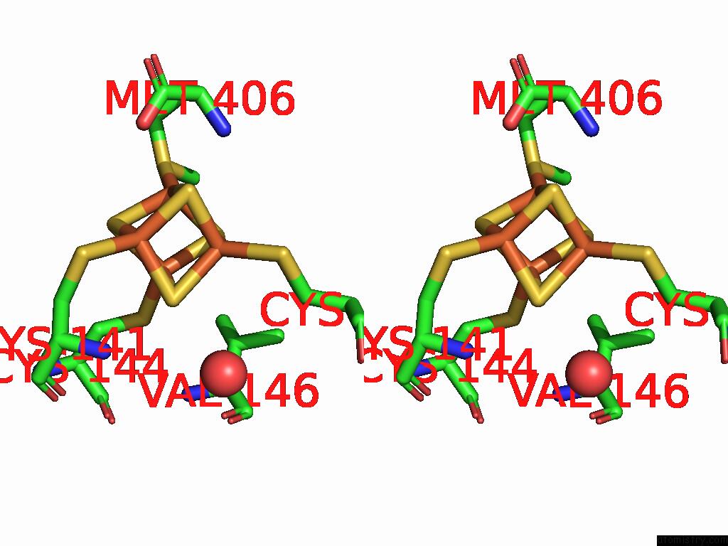
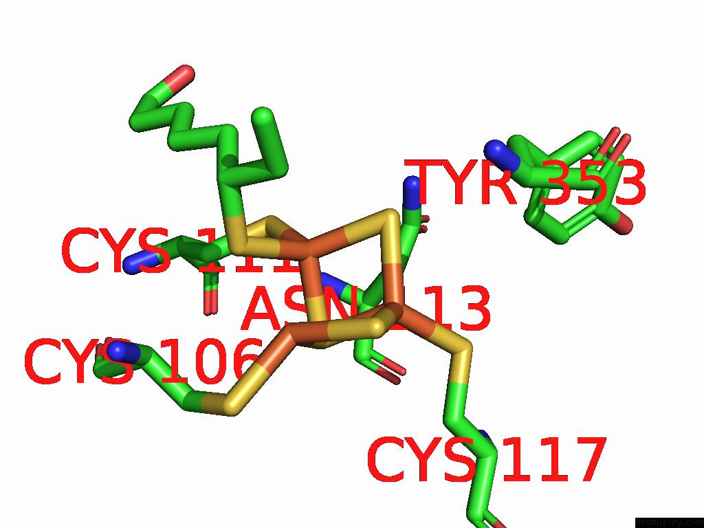
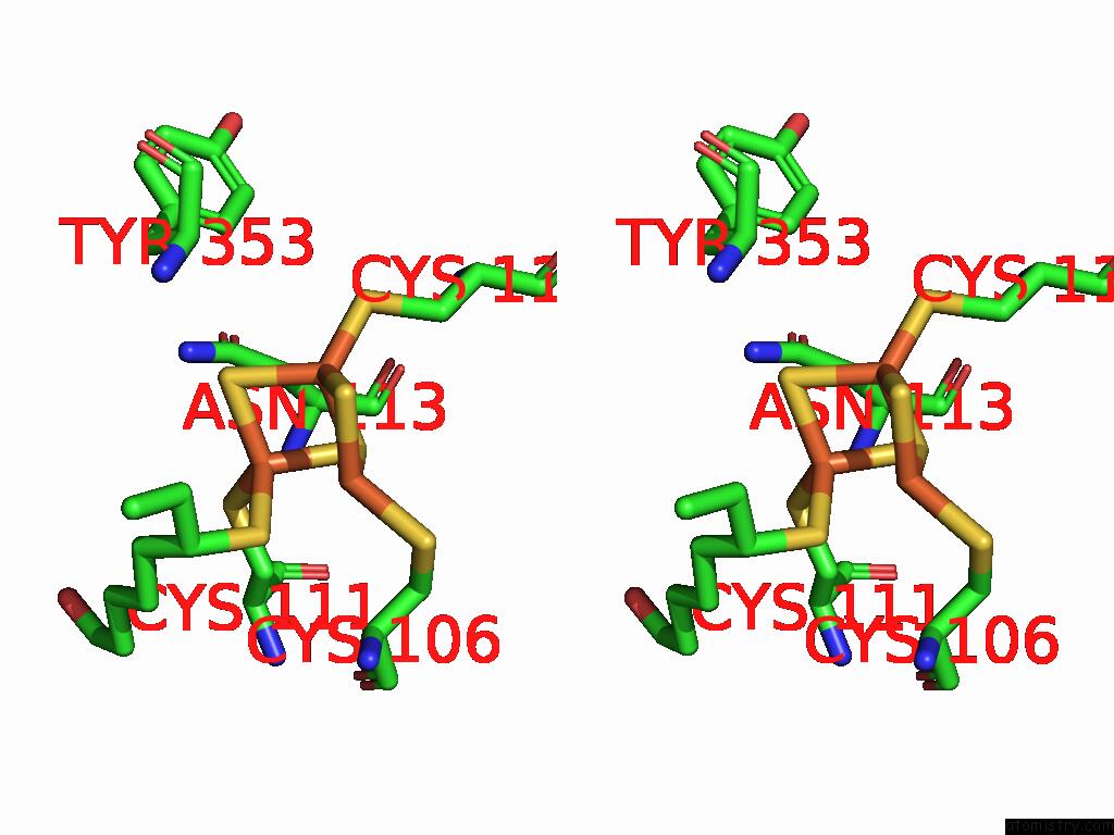
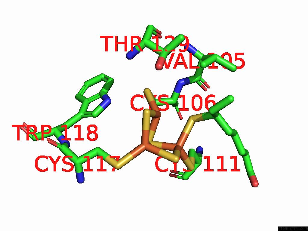
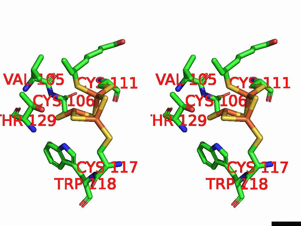
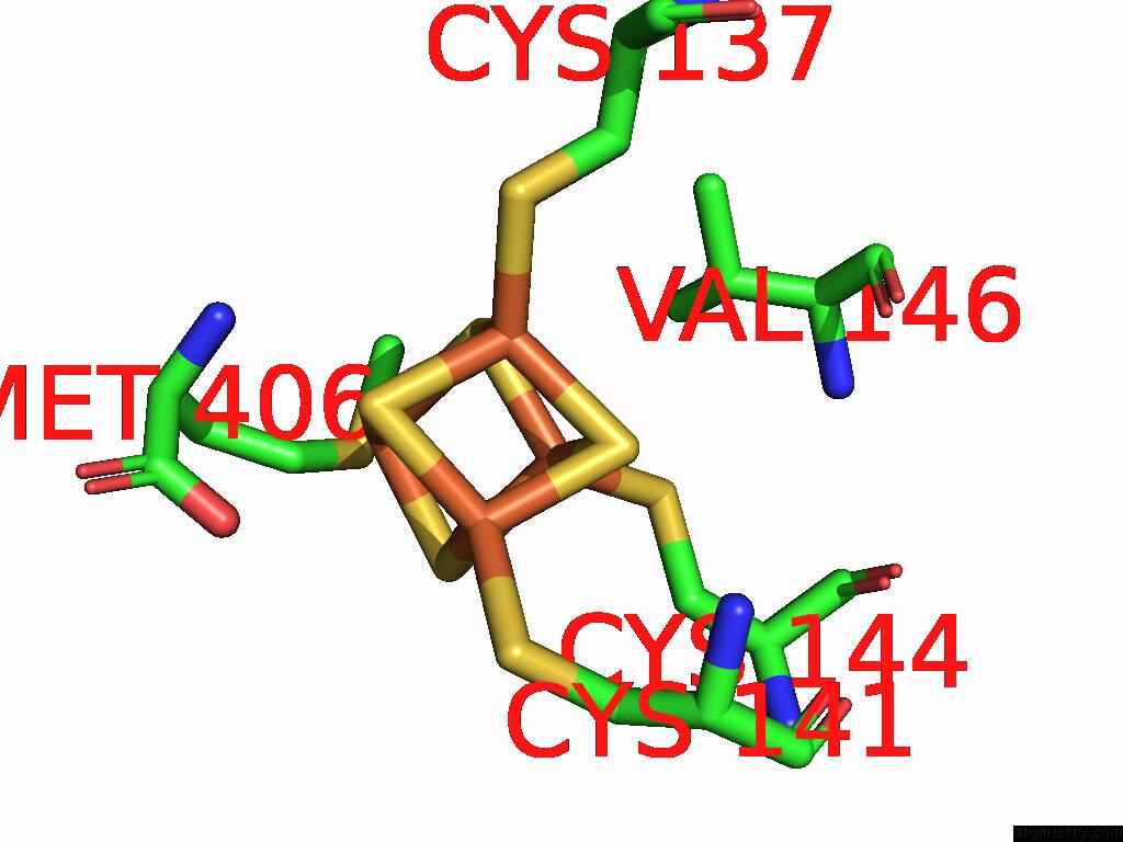
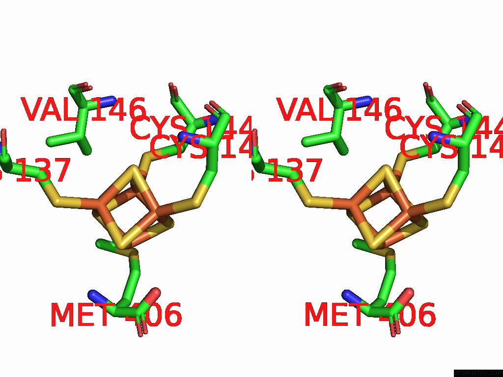
Iron binding site 1 out of 21 in 8ugo
Go back to
Iron binding site 1 out
of 21 in the Structure of the Complex Between Human Lias and H-Protein in the Presence of 5'-Deoxyadenosine
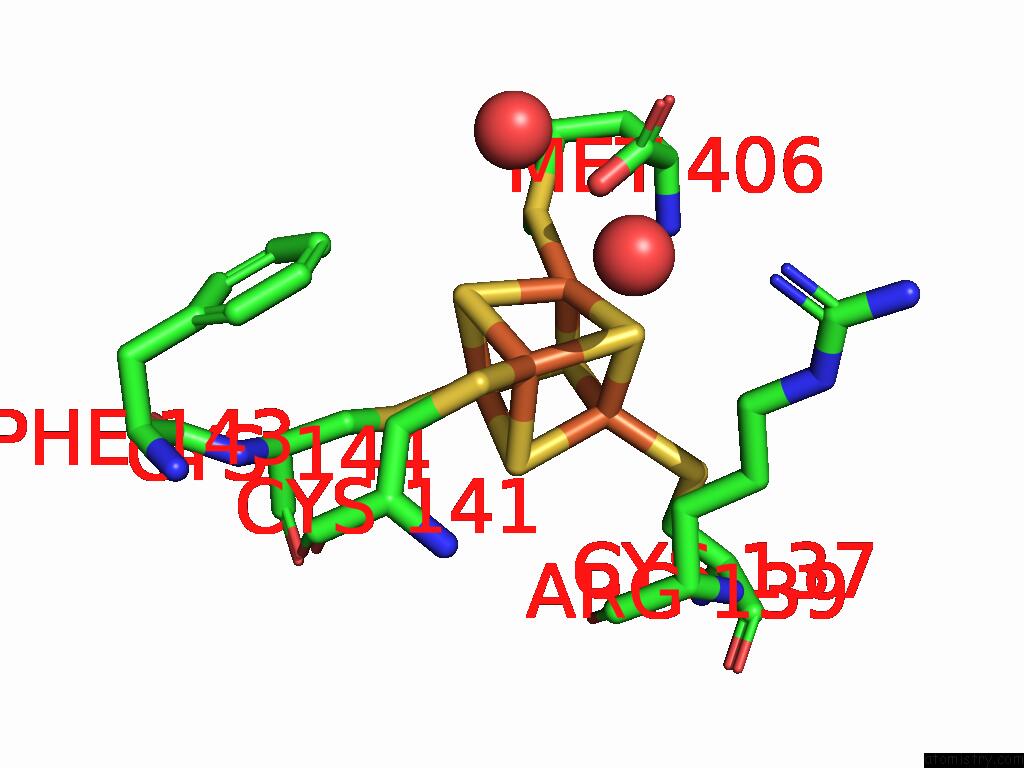
Mono view
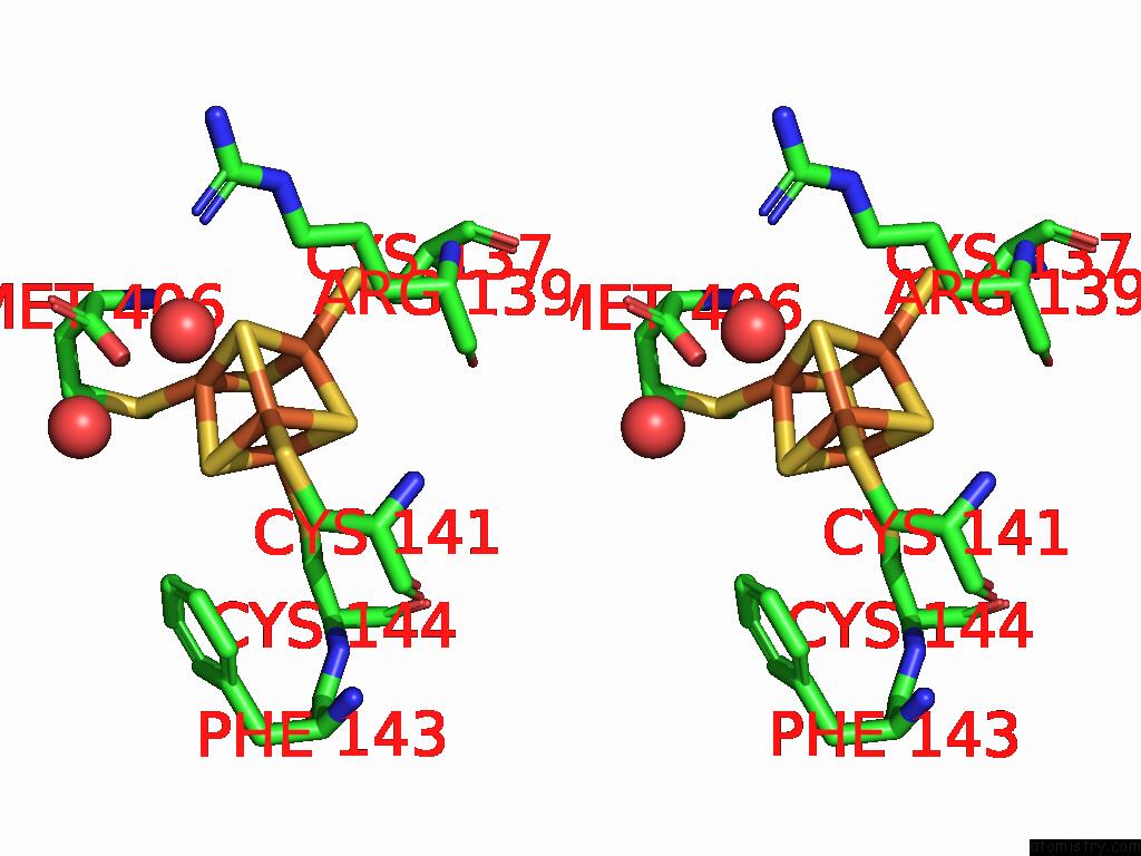
Stereo pair view
Mono view
Stereo pair view
A full contact list of Iron with other atoms in the Fe binding
site number 1 of Structure of the Complex Between Human Lias and H-Protein in the Presence of 5'-Deoxyadenosine within 5.0Å range:
|
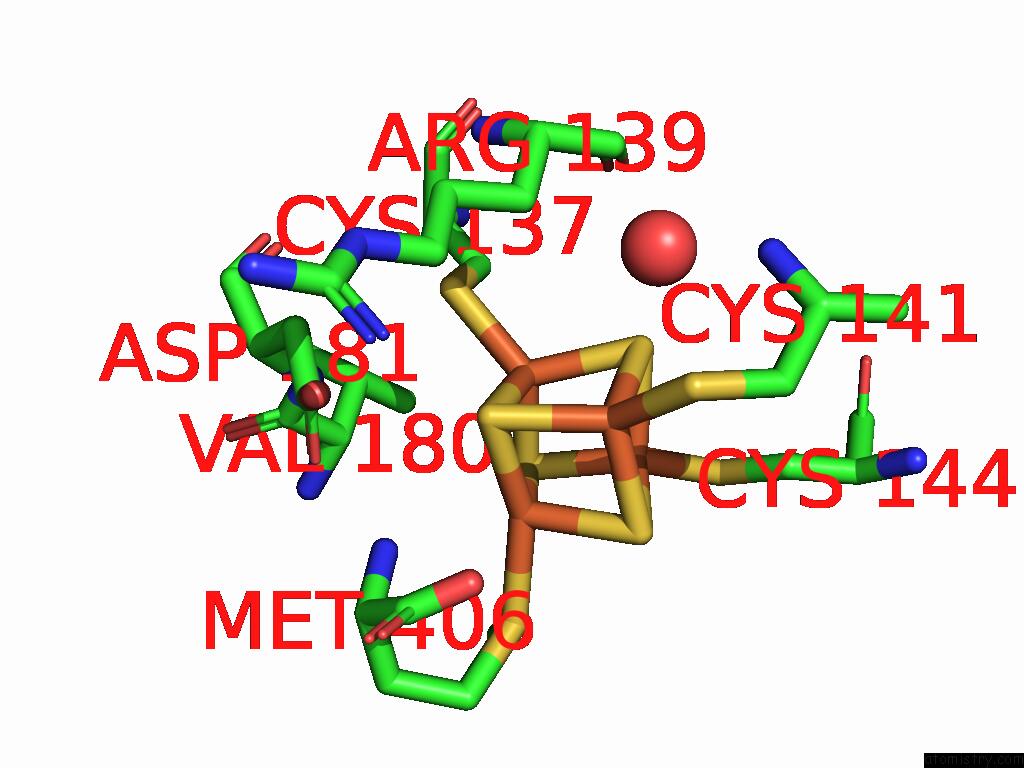
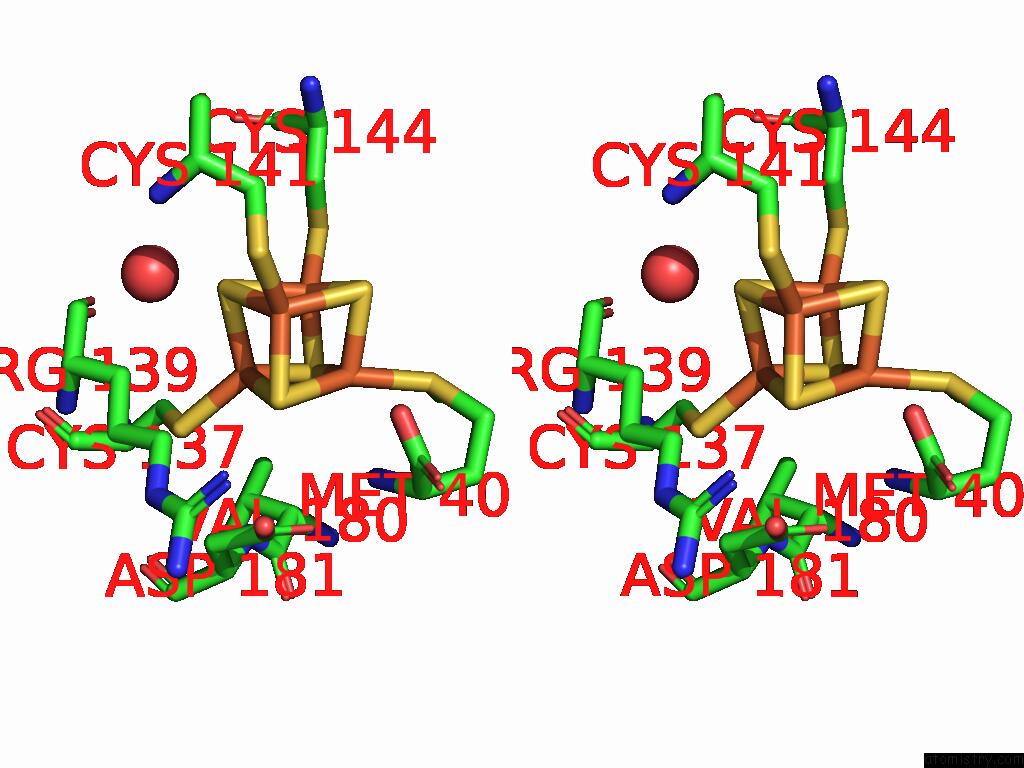
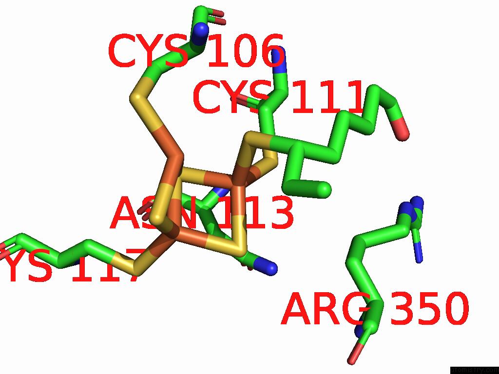
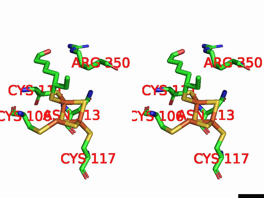
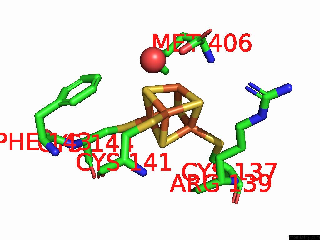
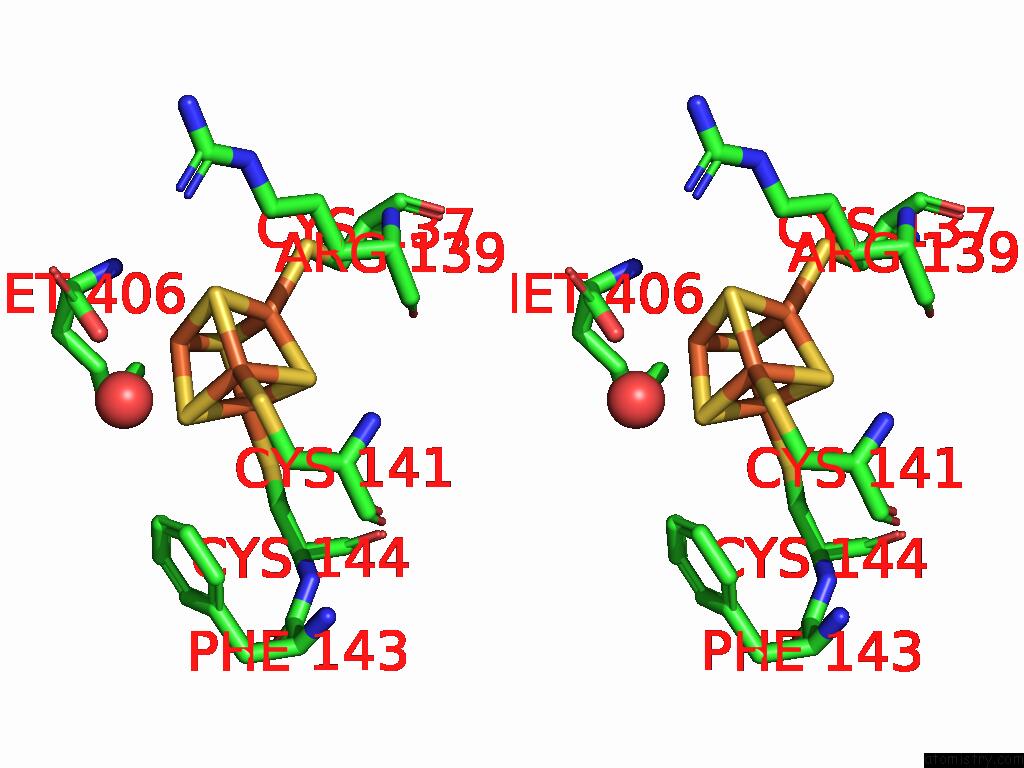
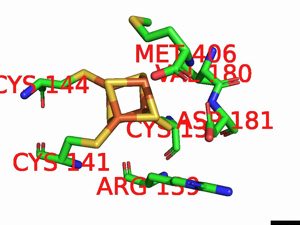
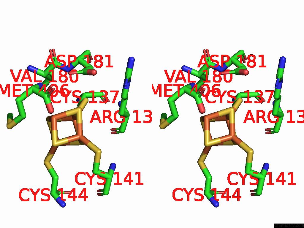
Iron binding site 2 out of 21 in 8ugo
Go back to
Iron binding site 2 out
of 21 in the Structure of the Complex Between Human Lias and H-Protein in the Presence of 5'-Deoxyadenosine
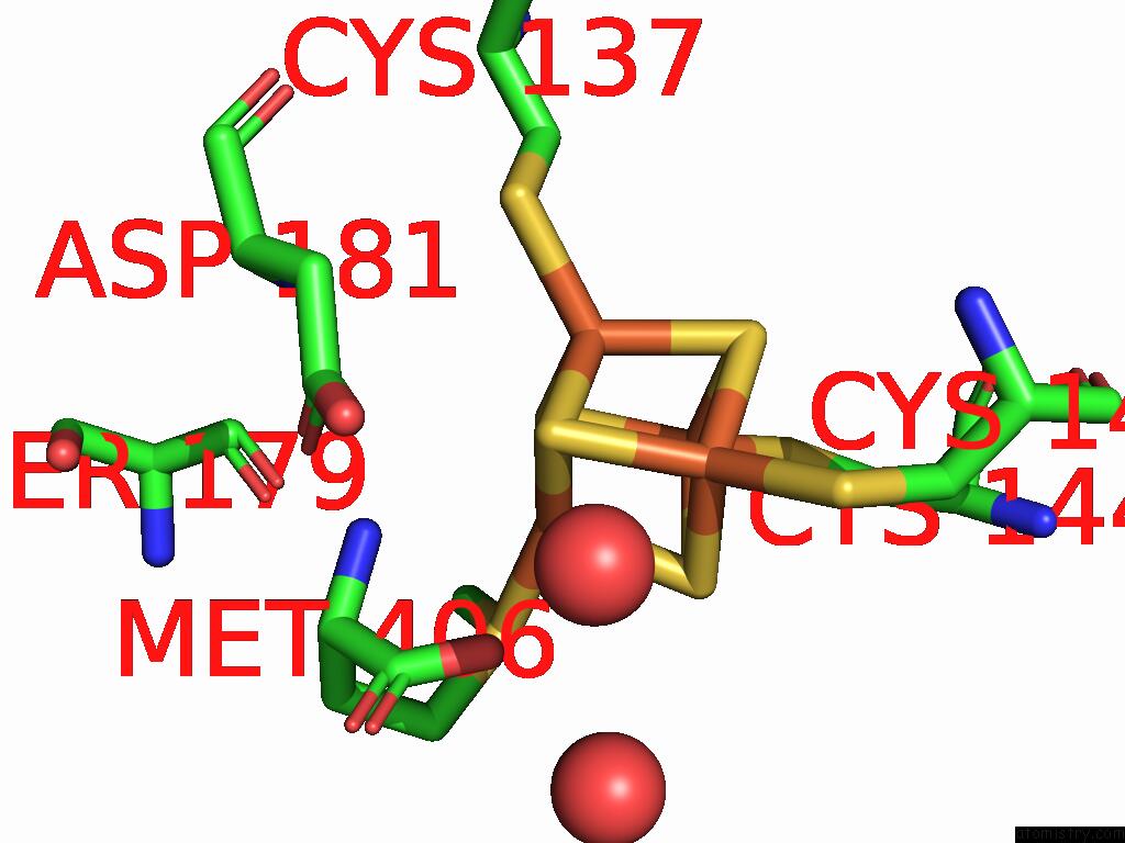
Mono view
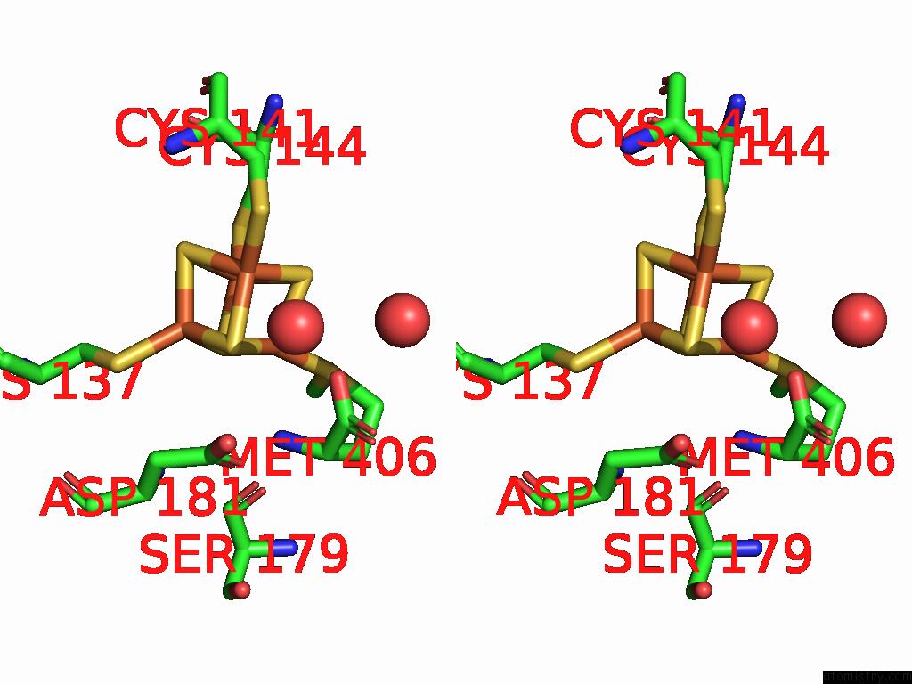
Stereo pair view
Mono view
Stereo pair view
A full contact list of Iron with other atoms in the Fe binding
site number 2 of Structure of the Complex Between Human Lias and H-Protein in the Presence of 5'-Deoxyadenosine within 5.0Å range:
|
Iron binding site 3 out of 21 in 8ugo
Go back to
Iron binding site 3 out
of 21 in the Structure of the Complex Between Human Lias and H-Protein in the Presence of 5'-Deoxyadenosine
Mono view
Stereo pair view
Mono view
Stereo pair view
A full contact list of Iron with other atoms in the Fe binding
site number 3 of Structure of the Complex Between Human Lias and H-Protein in the Presence of 5'-Deoxyadenosine within 5.0Å range:
|
Iron binding site 4 out of 21 in 8ugo
Go back to
Iron binding site 4 out
of 21 in the Structure of the Complex Between Human Lias and H-Protein in the Presence of 5'-Deoxyadenosine
Mono view
Stereo pair view
Mono view
Stereo pair view
A full contact list of Iron with other atoms in the Fe binding
site number 4 of Structure of the Complex Between Human Lias and H-Protein in the Presence of 5'-Deoxyadenosine within 5.0Å range:
|
Iron binding site 5 out of 21 in 8ugo
Go back to
Iron binding site 5 out
of 21 in the Structure of the Complex Between Human Lias and H-Protein in the Presence of 5'-Deoxyadenosine
Mono view
Stereo pair view
Mono view
Stereo pair view
A full contact list of Iron with other atoms in the Fe binding
site number 5 of Structure of the Complex Between Human Lias and H-Protein in the Presence of 5'-Deoxyadenosine within 5.0Å range:
|
Iron binding site 6 out of 21 in 8ugo
Go back to
Iron binding site 6 out
of 21 in the Structure of the Complex Between Human Lias and H-Protein in the Presence of 5'-Deoxyadenosine
Mono view
Stereo pair view
Mono view
Stereo pair view
A full contact list of Iron with other atoms in the Fe binding
site number 6 of Structure of the Complex Between Human Lias and H-Protein in the Presence of 5'-Deoxyadenosine within 5.0Å range:
|
Iron binding site 7 out of 21 in 8ugo
Go back to
Iron binding site 7 out
of 21 in the Structure of the Complex Between Human Lias and H-Protein in the Presence of 5'-Deoxyadenosine
Mono view
Stereo pair view
Mono view
Stereo pair view
A full contact list of Iron with other atoms in the Fe binding
site number 7 of Structure of the Complex Between Human Lias and H-Protein in the Presence of 5'-Deoxyadenosine within 5.0Å range:
|
Iron binding site 8 out of 21 in 8ugo
Go back to
Iron binding site 8 out
of 21 in the Structure of the Complex Between Human Lias and H-Protein in the Presence of 5'-Deoxyadenosine
Mono view
Stereo pair view
Mono view
Stereo pair view
A full contact list of Iron with other atoms in the Fe binding
site number 8 of Structure of the Complex Between Human Lias and H-Protein in the Presence of 5'-Deoxyadenosine within 5.0Å range:
|
Iron binding site 9 out of 21 in 8ugo
Go back to
Iron binding site 9 out
of 21 in the Structure of the Complex Between Human Lias and H-Protein in the Presence of 5'-Deoxyadenosine
Mono view
Stereo pair view
Mono view
Stereo pair view
A full contact list of Iron with other atoms in the Fe binding
site number 9 of Structure of the Complex Between Human Lias and H-Protein in the Presence of 5'-Deoxyadenosine within 5.0Å range:
|
Iron binding site 10 out of 21 in 8ugo
Go back to
Iron binding site 10 out
of 21 in the Structure of the Complex Between Human Lias and H-Protein in the Presence of 5'-Deoxyadenosine
Mono view
Stereo pair view
Mono view
Stereo pair view
A full contact list of Iron with other atoms in the Fe binding
site number 10 of Structure of the Complex Between Human Lias and H-Protein in the Presence of 5'-Deoxyadenosine within 5.0Å range:
|
Reference:
O.A.Esakova,
D.M.Warui,
S.S.Neti,
J.N.Alumasa,
S.J.Booker.
Structural Basis For the Mechanism of the Human Lipoyl Synthase (Lias) and Its Complex with the H-Protein To Be Published.
Page generated: Thu Aug 7 23:57:37 2025
Last articles
Y in 1DDEY in 1FS8
Y in 1FS7
Y in 1FS9
Xe in 7B9A
Xe in 9KUM
Xe in 9KUL
Xe in 9KUK
Xe in 9CS5
Xe in 7TSJ