Iron »
PDB 8yay-8zge »
8yhn »
Iron in PDB 8yhn: Crystal Structure of Cytochrome P450 107P2 From Streptomyces Avermitilis
Protein crystallography data
The structure of Crystal Structure of Cytochrome P450 107P2 From Streptomyces Avermitilis, PDB code: 8yhn
was solved by
E.S.Jeong,
V.C.Kim,
C.M.Kim,
Y.B.Lee,
with X-Ray Crystallography technique. A brief refinement statistics is given in the table below:
| Resolution Low / High (Å) | 44.21 / 1.99 |
| Space group | P 21 21 21 |
| Cell size a, b, c (Å), α, β, γ (°) | 59.128, 66.44, 111.756, 90, 90, 90 |
| R / Rfree (%) | 22.3 / 27.2 |
Iron Binding Sites:
The binding sites of Iron atom in the Crystal Structure of Cytochrome P450 107P2 From Streptomyces Avermitilis
(pdb code 8yhn). This binding sites where shown within
5.0 Angstroms radius around Iron atom.
In total only one binding site of Iron was determined in the Crystal Structure of Cytochrome P450 107P2 From Streptomyces Avermitilis, PDB code: 8yhn:
In total only one binding site of Iron was determined in the Crystal Structure of Cytochrome P450 107P2 From Streptomyces Avermitilis, PDB code: 8yhn:
Iron binding site 1 out of 1 in 8yhn
Go back to
Iron binding site 1 out
of 1 in the Crystal Structure of Cytochrome P450 107P2 From Streptomyces Avermitilis
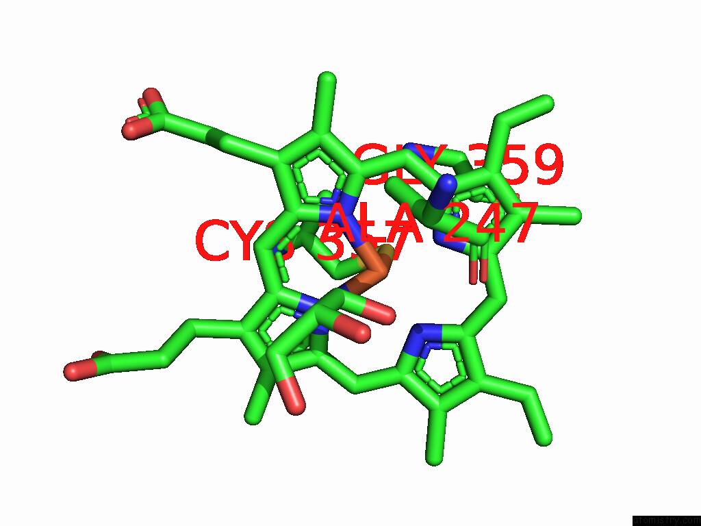
Mono view
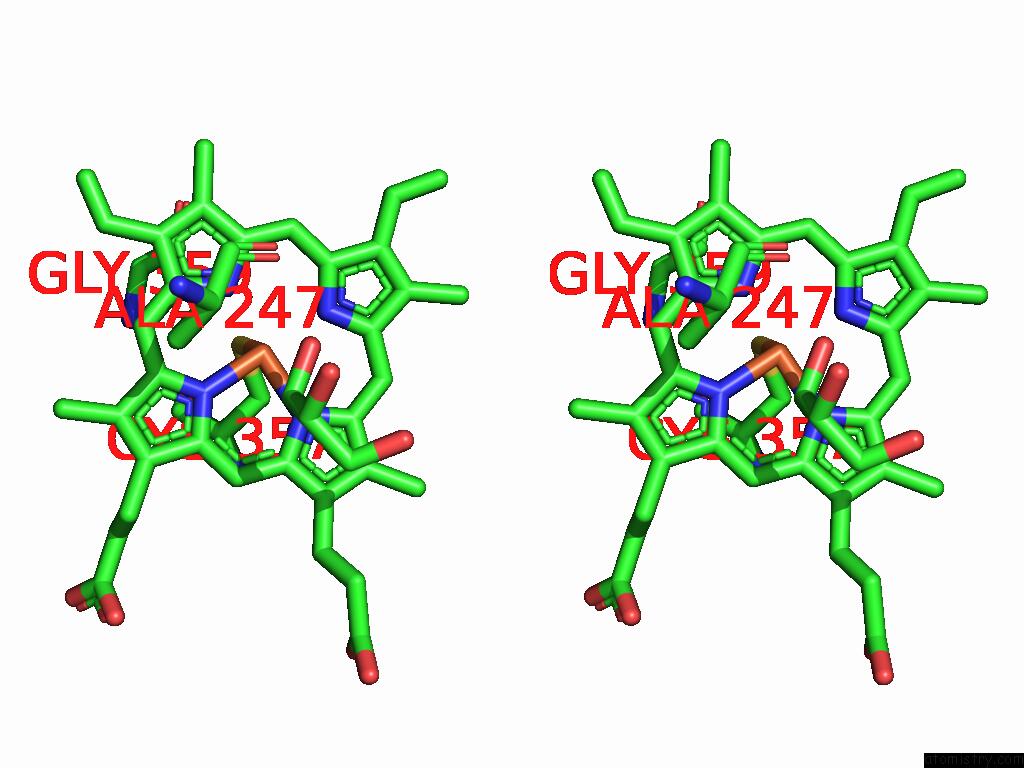
Stereo pair view
Mono view
Stereo pair view
A full contact list of Iron with other atoms in the Fe binding
site number 1 of Crystal Structure of Cytochrome P450 107P2 From Streptomyces Avermitilis within 5.0Å range:
|
Reference:
L.-H.Xu,
E.S.Jeong,
V.C.Kim,
C.M.Kim,
Y.B.Lee.
N/A N/A.
Page generated: Fri Aug 8 01:42:46 2025
Last articles
Zn in 1UXAZn in 1UX0
Zn in 1UWZ
Zn in 1UW0
Zn in 1UWY
Zn in 1UW1
Zn in 1UUP
Zn in 1UTZ
Zn in 1UV0
Zn in 1UVQ