Iron »
PDB 9e7h-9etz »
9eby »
Iron in PDB 9eby: Crystal Structure of Rufo in Complex with Mrylh
Protein crystallography data
The structure of Crystal Structure of Rufo in Complex with Mrylh, PDB code: 9eby
was solved by
K.Nolan,
Y.Wang,
with X-Ray Crystallography technique. A brief refinement statistics is given in the table below:
| Resolution Low / High (Å) | 45.28 / 2.03 |
| Space group | P 21 21 21 |
| Cell size a, b, c (Å), α, β, γ (°) | 55.163, 79.3, 88.511, 90, 90, 90 |
| R / Rfree (%) | 20.3 / 26.2 |
Iron Binding Sites:
The binding sites of Iron atom in the Crystal Structure of Rufo in Complex with Mrylh
(pdb code 9eby). This binding sites where shown within
5.0 Angstroms radius around Iron atom.
In total only one binding site of Iron was determined in the Crystal Structure of Rufo in Complex with Mrylh, PDB code: 9eby:
In total only one binding site of Iron was determined in the Crystal Structure of Rufo in Complex with Mrylh, PDB code: 9eby:
Iron binding site 1 out of 1 in 9eby
Go back to
Iron binding site 1 out
of 1 in the Crystal Structure of Rufo in Complex with Mrylh
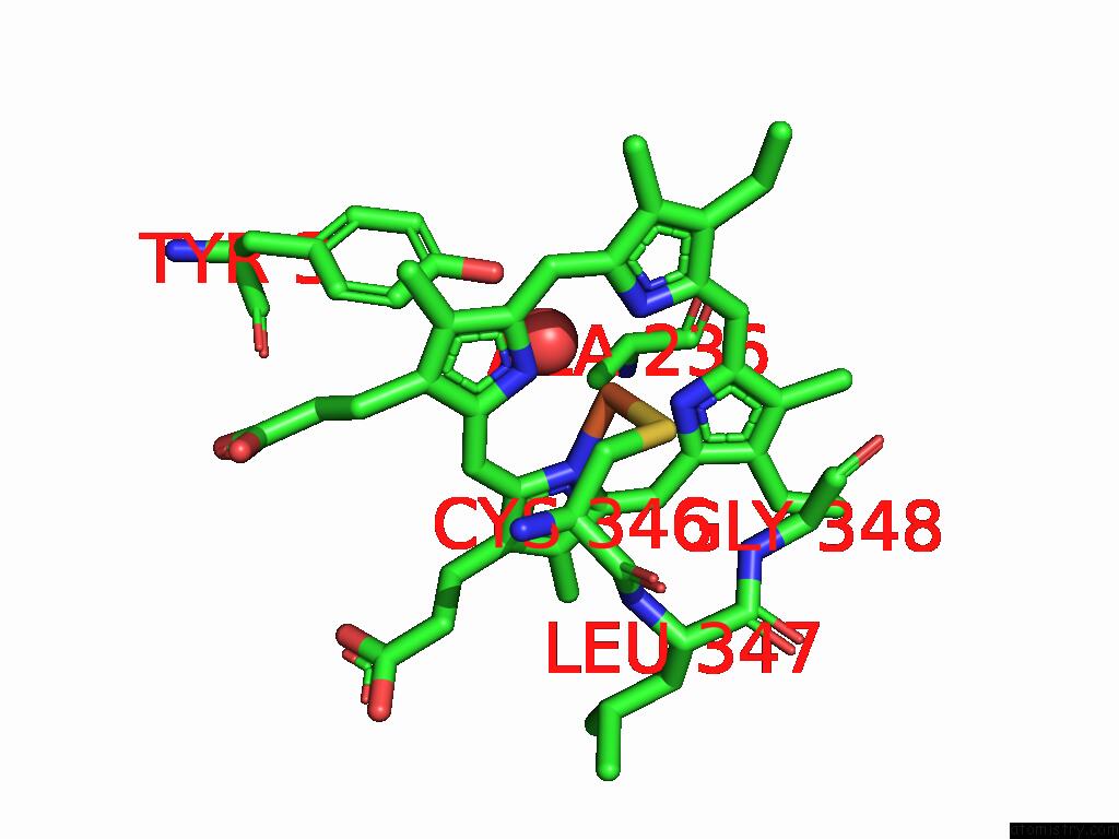
Mono view
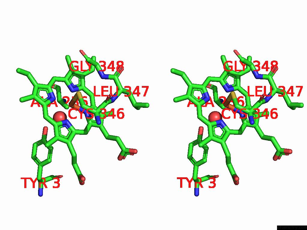
Stereo pair view
Mono view
Stereo pair view
A full contact list of Iron with other atoms in the Fe binding
site number 1 of Crystal Structure of Rufo in Complex with Mrylh within 5.0Å range:
|
Reference:
K.Nolan,
R.Usai,
B.Li,
S.Jordan,
Y.Wang.
Molecular Basis For Peptide Nitration By A Novel Cytochrome P450 Enzyme in Ripp Biosynthesis Acs Catalysis 10391 2025.
ISSN: ESSN 2155-5435
DOI: 10.1021/ACSCATAL.5C01932
Page generated: Fri Aug 8 04:08:25 2025
ISSN: ESSN 2155-5435
DOI: 10.1021/ACSCATAL.5C01932
Last articles
K in 3USZK in 3UM7
K in 3UMO
K in 3UMB
K in 3UGP
K in 3UGQ
K in 3UGO
K in 3UGX
K in 3TZE
K in 3U81