Iron »
PDB 9gz0-9imn »
9iki »
Iron in PDB 9iki: Bovine Heart Cytochrome C Oxidase in the Nitrous Oxide-Bound Fully Reduced State
Enzymatic activity of Bovine Heart Cytochrome C Oxidase in the Nitrous Oxide-Bound Fully Reduced State
All present enzymatic activity of Bovine Heart Cytochrome C Oxidase in the Nitrous Oxide-Bound Fully Reduced State:
7.1.1.9;
7.1.1.9;
Protein crystallography data
The structure of Bovine Heart Cytochrome C Oxidase in the Nitrous Oxide-Bound Fully Reduced State, PDB code: 9iki
was solved by
K.Muramoto,
T.Ide,
K.Shinzawa-Itoh,
with X-Ray Crystallography technique. A brief refinement statistics is given in the table below:
| Resolution Low / High (Å) | 40.00 / 1.75 |
| Space group | P 21 21 21 |
| Cell size a, b, c (Å), α, β, γ (°) | 181.9, 204, 177.7, 90, 90, 90 |
| R / Rfree (%) | 15.3 / 18.9 |
Other elements in 9iki:
The structure of Bovine Heart Cytochrome C Oxidase in the Nitrous Oxide-Bound Fully Reduced State also contains other interesting chemical elements:
| Magnesium | (Mg) | 2 atoms |
| Zinc | (Zn) | 2 atoms |
| Copper | (Cu) | 6 atoms |
| Sodium | (Na) | 2 atoms |
Iron Binding Sites:
The binding sites of Iron atom in the Bovine Heart Cytochrome C Oxidase in the Nitrous Oxide-Bound Fully Reduced State
(pdb code 9iki). This binding sites where shown within
5.0 Angstroms radius around Iron atom.
In total 4 binding sites of Iron where determined in the Bovine Heart Cytochrome C Oxidase in the Nitrous Oxide-Bound Fully Reduced State, PDB code: 9iki:
Jump to Iron binding site number: 1; 2; 3; 4;
In total 4 binding sites of Iron where determined in the Bovine Heart Cytochrome C Oxidase in the Nitrous Oxide-Bound Fully Reduced State, PDB code: 9iki:
Jump to Iron binding site number: 1; 2; 3; 4;
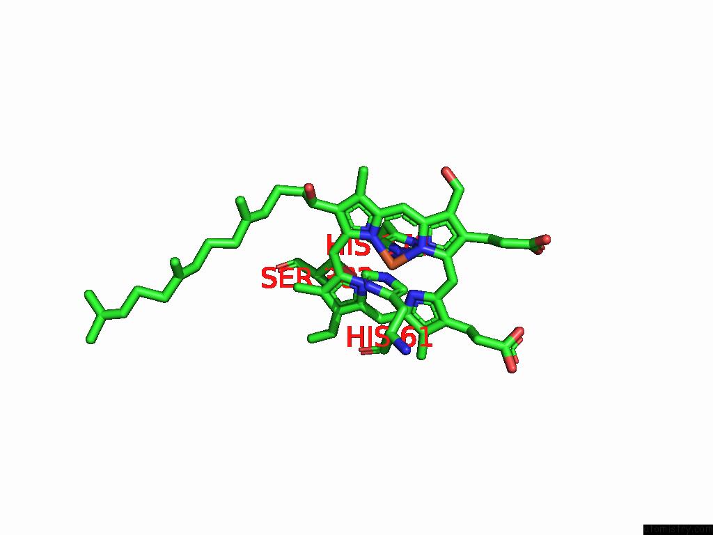
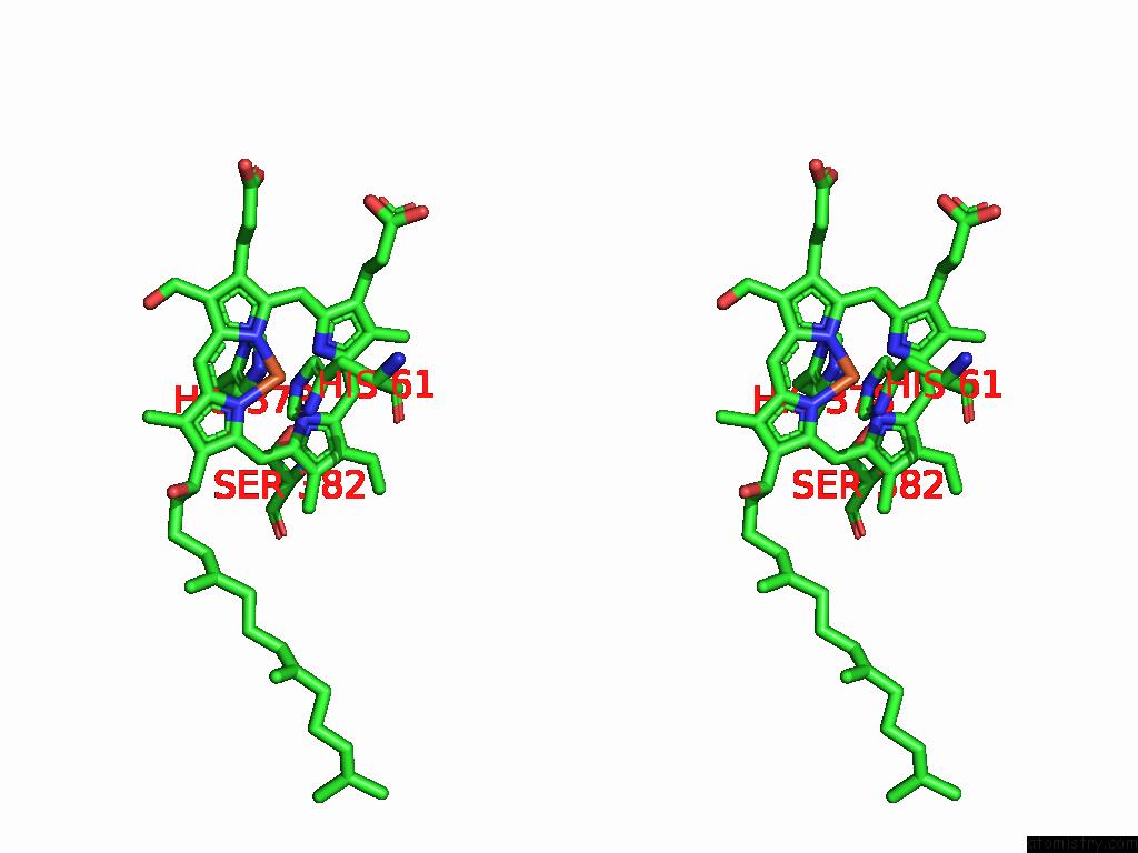
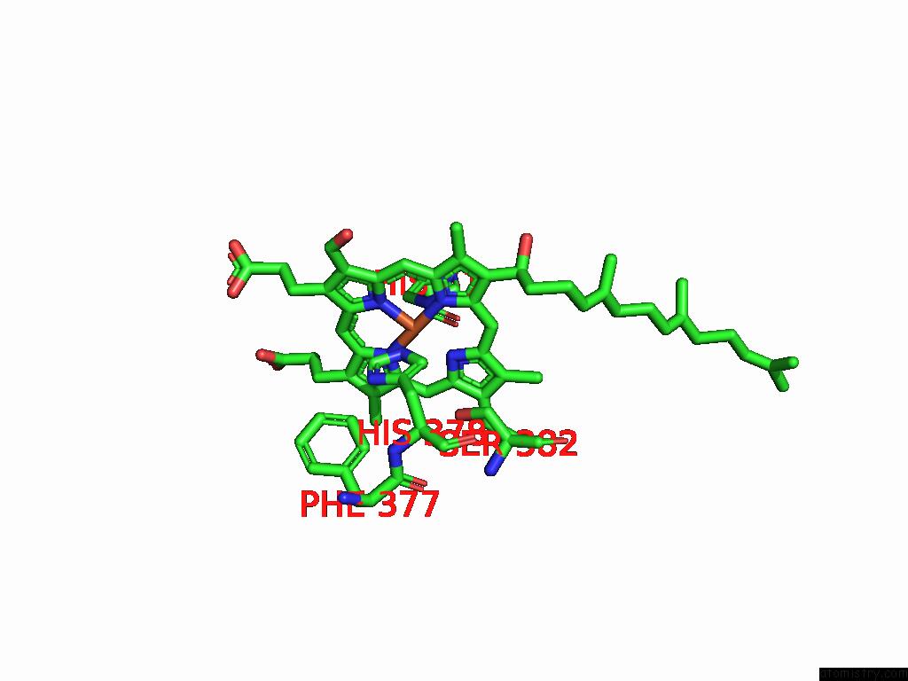
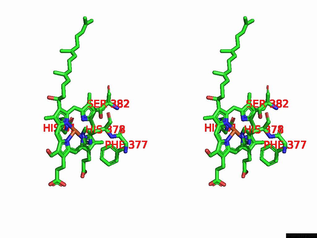
Iron binding site 1 out of 4 in 9iki
Go back to
Iron binding site 1 out
of 4 in the Bovine Heart Cytochrome C Oxidase in the Nitrous Oxide-Bound Fully Reduced State
Mono view
Stereo pair view
Mono view
Stereo pair view
A full contact list of Iron with other atoms in the Fe binding
site number 1 of Bovine Heart Cytochrome C Oxidase in the Nitrous Oxide-Bound Fully Reduced State within 5.0Å range:
|
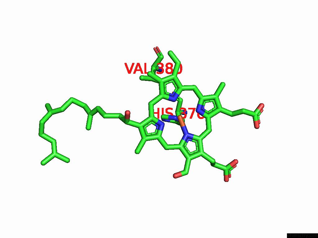
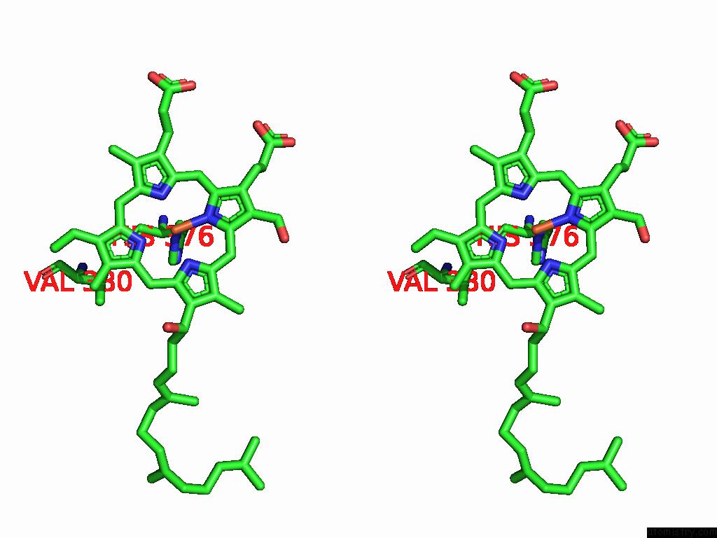
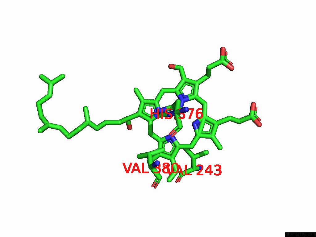
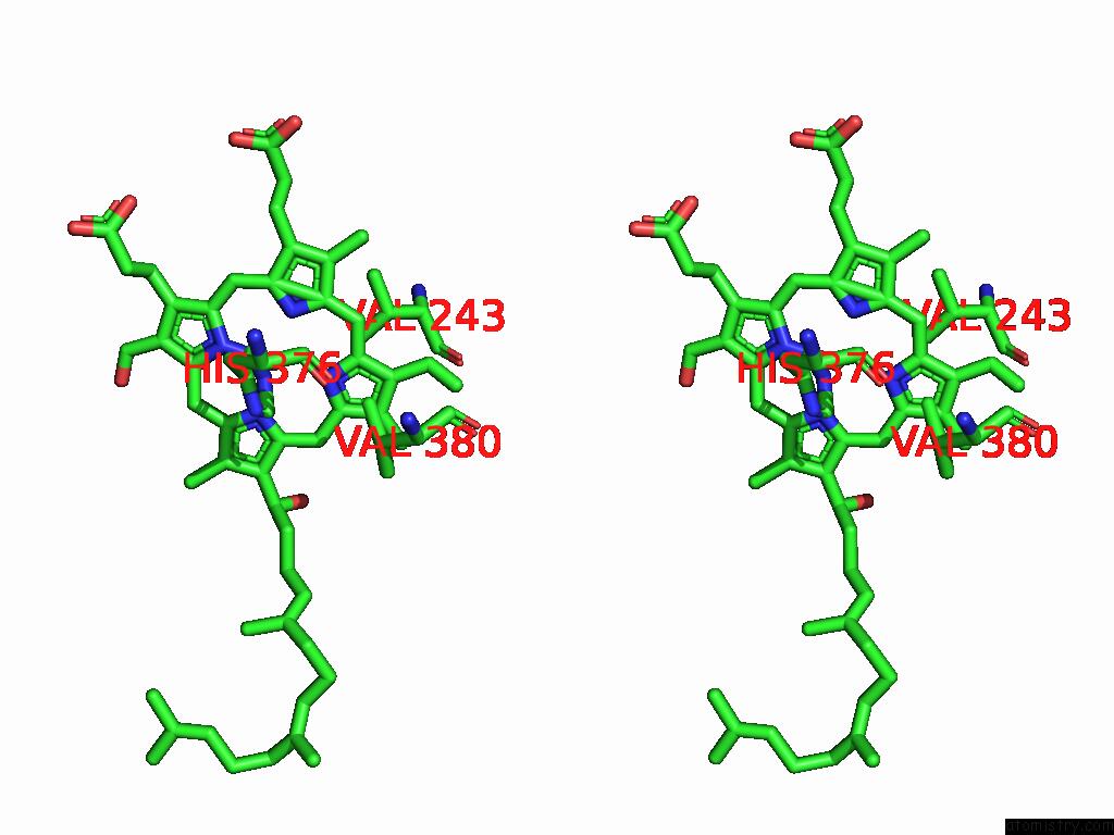
Iron binding site 2 out of 4 in 9iki
Go back to
Iron binding site 2 out
of 4 in the Bovine Heart Cytochrome C Oxidase in the Nitrous Oxide-Bound Fully Reduced State
Mono view
Stereo pair view
Mono view
Stereo pair view
A full contact list of Iron with other atoms in the Fe binding
site number 2 of Bovine Heart Cytochrome C Oxidase in the Nitrous Oxide-Bound Fully Reduced State within 5.0Å range:
|
Iron binding site 3 out of 4 in 9iki
Go back to
Iron binding site 3 out
of 4 in the Bovine Heart Cytochrome C Oxidase in the Nitrous Oxide-Bound Fully Reduced State
Mono view
Stereo pair view
Mono view
Stereo pair view
A full contact list of Iron with other atoms in the Fe binding
site number 3 of Bovine Heart Cytochrome C Oxidase in the Nitrous Oxide-Bound Fully Reduced State within 5.0Å range:
|
Iron binding site 4 out of 4 in 9iki
Go back to
Iron binding site 4 out
of 4 in the Bovine Heart Cytochrome C Oxidase in the Nitrous Oxide-Bound Fully Reduced State
Mono view
Stereo pair view
Mono view
Stereo pair view
A full contact list of Iron with other atoms in the Fe binding
site number 4 of Bovine Heart Cytochrome C Oxidase in the Nitrous Oxide-Bound Fully Reduced State within 5.0Å range:
|
Reference:
K.Muramoto,
T.Ide,
K.Shinzawa-Itoh.
The Binding Sites of Carbon Dioxide, Nitrous Oxide, and Xenon Reveal A Putative Exhaust Channel For Bovine Cytochrome C Oxidase. J.Biol.Chem. 10395 2025.
ISSN: ESSN 1083-351X
PubMed: 40543594
DOI: 10.1016/J.JBC.2025.110395
Page generated: Fri Aug 8 06:50:49 2025
ISSN: ESSN 1083-351X
PubMed: 40543594
DOI: 10.1016/J.JBC.2025.110395
Last articles
Hg in 1LUGHg in 1KDG
Hg in 1L8D
Hg in 1L9A
Hg in 1KZL
Hg in 1KWQ
Hg in 1JQC
Hg in 1KGZ
Hg in 1K1E
Hg in 1JPR