Iron »
PDB 9kqb-9n5u »
9kt5 »
Iron in PDB 9kt5: Synchrotron X-Ray Crystal Structure of Oxygen-Bound F87A/F393H P450BM3 with Decoy C7PROPHE (N-Enanthyl-L-Prolyl-L-Phenylalanine) and Substrate Styrene at 2 Mgy X-Ray Dose
Enzymatic activity of Synchrotron X-Ray Crystal Structure of Oxygen-Bound F87A/F393H P450BM3 with Decoy C7PROPHE (N-Enanthyl-L-Prolyl-L-Phenylalanine) and Substrate Styrene at 2 Mgy X-Ray Dose
All present enzymatic activity of Synchrotron X-Ray Crystal Structure of Oxygen-Bound F87A/F393H P450BM3 with Decoy C7PROPHE (N-Enanthyl-L-Prolyl-L-Phenylalanine) and Substrate Styrene at 2 Mgy X-Ray Dose:
1.14.14.1; 1.6.2.4;
1.14.14.1; 1.6.2.4;
Protein crystallography data
The structure of Synchrotron X-Ray Crystal Structure of Oxygen-Bound F87A/F393H P450BM3 with Decoy C7PROPHE (N-Enanthyl-L-Prolyl-L-Phenylalanine) and Substrate Styrene at 2 Mgy X-Ray Dose, PDB code: 9kt5
was solved by
S.Nagao,
W.Kuwano,
T.Tosha,
K.Yamashita,
J.K.Stanfield,
C.Kasai,
S.Ariyasu,
O.Shoji,
H.Sugimoto,
M.Kubo,
with X-Ray Crystallography technique. A brief refinement statistics is given in the table below:
| Resolution Low / High (Å) | 43.64 / 1.60 |
| Space group | P 21 21 21 |
| Cell size a, b, c (Å), α, β, γ (°) | 59.13, 129.34, 150.15, 90, 90, 90 |
| R / Rfree (%) | 14.9 / 20 |
Iron Binding Sites:
The binding sites of Iron atom in the Synchrotron X-Ray Crystal Structure of Oxygen-Bound F87A/F393H P450BM3 with Decoy C7PROPHE (N-Enanthyl-L-Prolyl-L-Phenylalanine) and Substrate Styrene at 2 Mgy X-Ray Dose
(pdb code 9kt5). This binding sites where shown within
5.0 Angstroms radius around Iron atom.
In total 4 binding sites of Iron where determined in the Synchrotron X-Ray Crystal Structure of Oxygen-Bound F87A/F393H P450BM3 with Decoy C7PROPHE (N-Enanthyl-L-Prolyl-L-Phenylalanine) and Substrate Styrene at 2 Mgy X-Ray Dose, PDB code: 9kt5:
Jump to Iron binding site number: 1; 2; 3; 4;
In total 4 binding sites of Iron where determined in the Synchrotron X-Ray Crystal Structure of Oxygen-Bound F87A/F393H P450BM3 with Decoy C7PROPHE (N-Enanthyl-L-Prolyl-L-Phenylalanine) and Substrate Styrene at 2 Mgy X-Ray Dose, PDB code: 9kt5:
Jump to Iron binding site number: 1; 2; 3; 4;
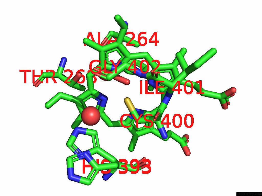
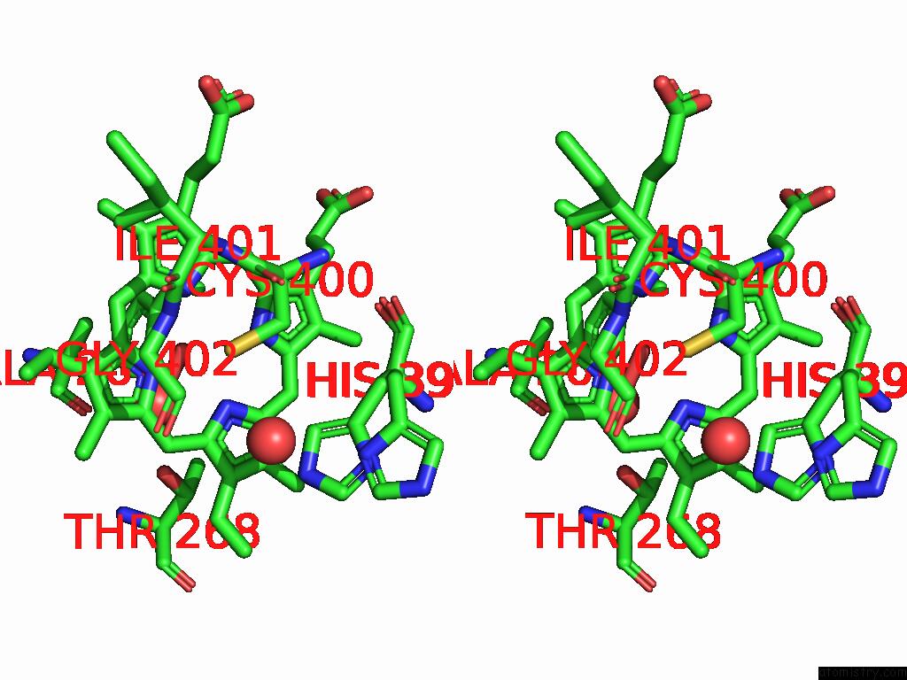
Iron binding site 1 out of 4 in 9kt5
Go back to
Iron binding site 1 out
of 4 in the Synchrotron X-Ray Crystal Structure of Oxygen-Bound F87A/F393H P450BM3 with Decoy C7PROPHE (N-Enanthyl-L-Prolyl-L-Phenylalanine) and Substrate Styrene at 2 Mgy X-Ray Dose
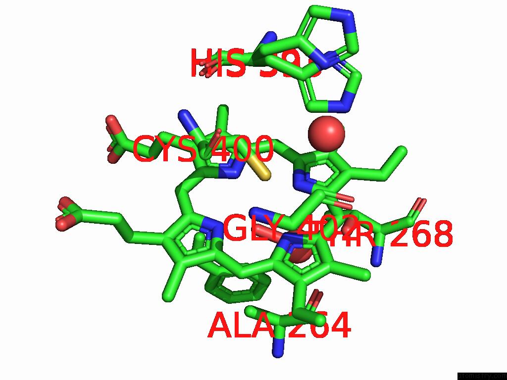
Mono view
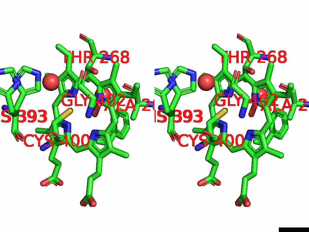
Stereo pair view
Mono view
Stereo pair view
A full contact list of Iron with other atoms in the Fe binding
site number 1 of Synchrotron X-Ray Crystal Structure of Oxygen-Bound F87A/F393H P450BM3 with Decoy C7PROPHE (N-Enanthyl-L-Prolyl-L-Phenylalanine) and Substrate Styrene at 2 Mgy X-Ray Dose within 5.0Å range:
|
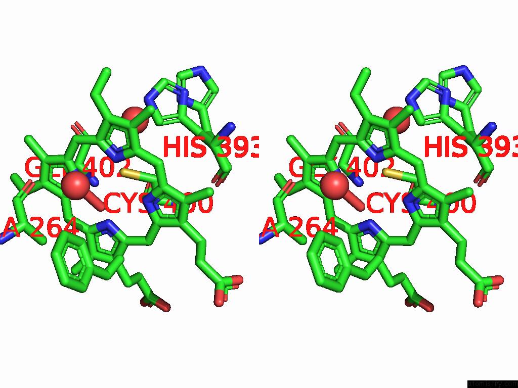
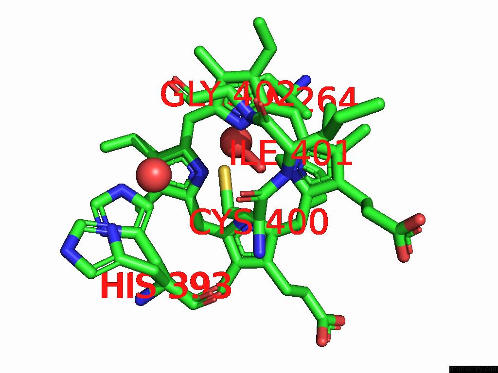
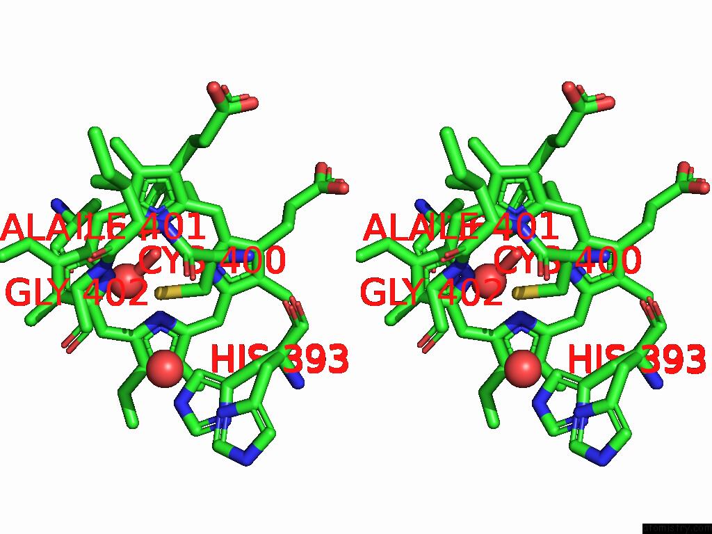
Iron binding site 2 out of 4 in 9kt5
Go back to
Iron binding site 2 out
of 4 in the Synchrotron X-Ray Crystal Structure of Oxygen-Bound F87A/F393H P450BM3 with Decoy C7PROPHE (N-Enanthyl-L-Prolyl-L-Phenylalanine) and Substrate Styrene at 2 Mgy X-Ray Dose
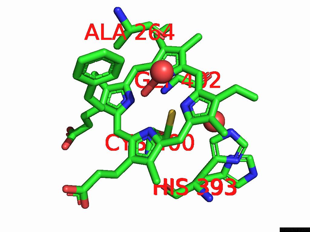
Mono view
Stereo pair view
Mono view
Stereo pair view
A full contact list of Iron with other atoms in the Fe binding
site number 2 of Synchrotron X-Ray Crystal Structure of Oxygen-Bound F87A/F393H P450BM3 with Decoy C7PROPHE (N-Enanthyl-L-Prolyl-L-Phenylalanine) and Substrate Styrene at 2 Mgy X-Ray Dose within 5.0Å range:
|
Iron binding site 3 out of 4 in 9kt5
Go back to
Iron binding site 3 out
of 4 in the Synchrotron X-Ray Crystal Structure of Oxygen-Bound F87A/F393H P450BM3 with Decoy C7PROPHE (N-Enanthyl-L-Prolyl-L-Phenylalanine) and Substrate Styrene at 2 Mgy X-Ray Dose
Mono view
Stereo pair view
Mono view
Stereo pair view
A full contact list of Iron with other atoms in the Fe binding
site number 3 of Synchrotron X-Ray Crystal Structure of Oxygen-Bound F87A/F393H P450BM3 with Decoy C7PROPHE (N-Enanthyl-L-Prolyl-L-Phenylalanine) and Substrate Styrene at 2 Mgy X-Ray Dose within 5.0Å range:
|
Iron binding site 4 out of 4 in 9kt5
Go back to
Iron binding site 4 out
of 4 in the Synchrotron X-Ray Crystal Structure of Oxygen-Bound F87A/F393H P450BM3 with Decoy C7PROPHE (N-Enanthyl-L-Prolyl-L-Phenylalanine) and Substrate Styrene at 2 Mgy X-Ray Dose
Mono view
Stereo pair view
Mono view
Stereo pair view
A full contact list of Iron with other atoms in the Fe binding
site number 4 of Synchrotron X-Ray Crystal Structure of Oxygen-Bound F87A/F393H P450BM3 with Decoy C7PROPHE (N-Enanthyl-L-Prolyl-L-Phenylalanine) and Substrate Styrene at 2 Mgy X-Ray Dose within 5.0Å range:
|
Reference:
S.Nagao,
W.Kuwano,
T.Tosha,
K.Yamashita,
J.K.Stanfield,
C.Kasai,
S.Ariyasu,
K.Hirata,
G.Ueno,
H.Murakami,
H.Ago,
M.Yamamoto,
O.Shoji,
H.Sugimoto,
M.Kubo.
Xfel Crystallography Reveals Catalytic Cycle Dynamics During Non-Native Substrate Oxidation By Cytochrome P450BM3. Commun Chem V. 8 63 2025.
ISSN: ESSN 2399-3669
PubMed: 40075209
DOI: 10.1038/S42004-025-01440-2
Page generated: Fri Aug 8 07:06:38 2025
ISSN: ESSN 2399-3669
PubMed: 40075209
DOI: 10.1038/S42004-025-01440-2
Last articles
Mg in 1VQ9Mg in 1VQ7
Mg in 1VQ5
Mg in 1VQ6
Mg in 1VQ8
Mg in 1VQ4
Mg in 1VPA
Mg in 1VPE
Mg in 1VOM
Mg in 1VMA