Iron »
PDB 9n5v-9vie »
9vic »
Iron in PDB 9vic: Cryo-Em Structure of Human Haemoglobin in the R Conformation
Iron Binding Sites:
The binding sites of Iron atom in the Cryo-Em Structure of Human Haemoglobin in the R Conformation
(pdb code 9vic). This binding sites where shown within
5.0 Angstroms radius around Iron atom.
In total 2 binding sites of Iron where determined in the Cryo-Em Structure of Human Haemoglobin in the R Conformation, PDB code: 9vic:
Jump to Iron binding site number: 1; 2;
In total 2 binding sites of Iron where determined in the Cryo-Em Structure of Human Haemoglobin in the R Conformation, PDB code: 9vic:
Jump to Iron binding site number: 1; 2;
Iron binding site 1 out of 2 in 9vic
Go back to
Iron binding site 1 out
of 2 in the Cryo-Em Structure of Human Haemoglobin in the R Conformation
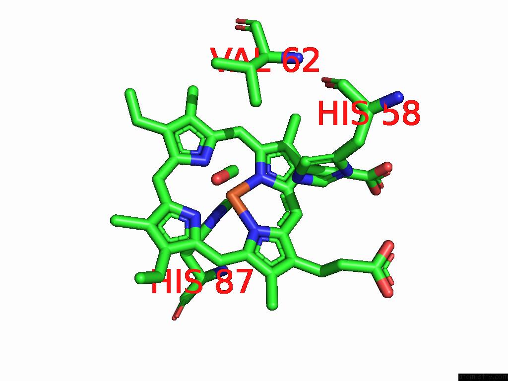
Mono view
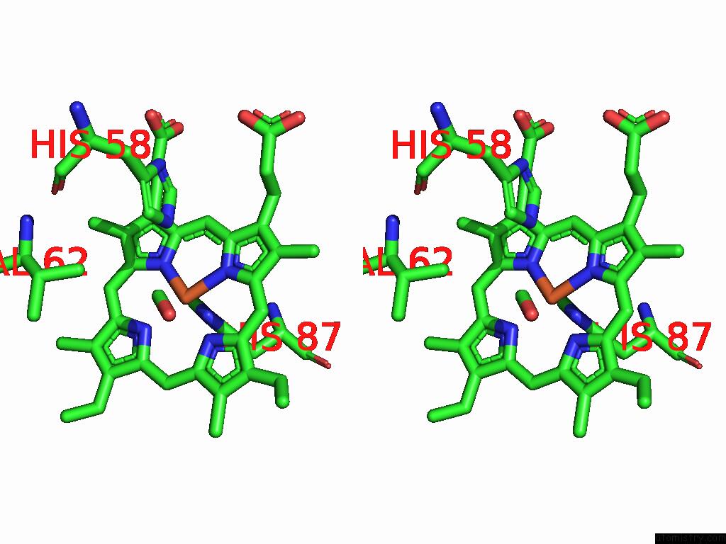
Stereo pair view
Mono view
Stereo pair view
A full contact list of Iron with other atoms in the Fe binding
site number 1 of Cryo-Em Structure of Human Haemoglobin in the R Conformation within 5.0Å range:
|
Iron binding site 2 out of 2 in 9vic
Go back to
Iron binding site 2 out
of 2 in the Cryo-Em Structure of Human Haemoglobin in the R Conformation
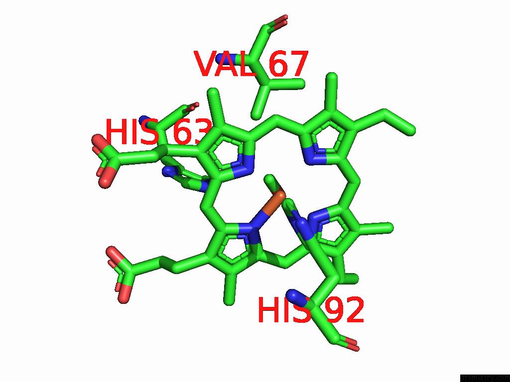
Mono view
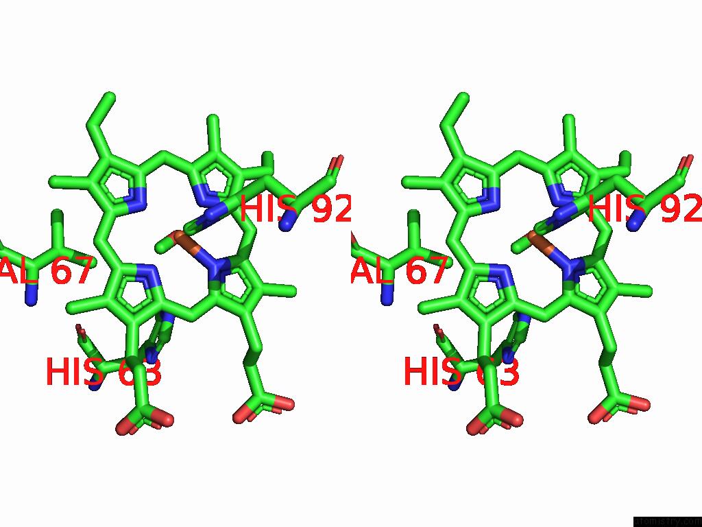
Stereo pair view
Mono view
Stereo pair view
A full contact list of Iron with other atoms in the Fe binding
site number 2 of Cryo-Em Structure of Human Haemoglobin in the R Conformation within 5.0Å range:
|
Reference:
K.Takahashi,
Y.Lee,
T.Nishizawa,
J.R.H.Tame.
Conformational Analysis of Liganded Human Hemoglobin By Cryo Electron Microscopy Biorxiv 2025.
ISSN: ISSN 2692-8205
DOI: 10.1101/2025.07.07.661630
Page generated: Fri Aug 8 08:22:19 2025
ISSN: ISSN 2692-8205
DOI: 10.1101/2025.07.07.661630
Last articles
K in 3PI7K in 3PEI
K in 3P6T
K in 3P6P
K in 3P6O
K in 3P6N
K in 3P1F
K in 3P1V
K in 3P6M
K in 3P1E