Iron »
PDB 101m-1a8f »
1a3o »
Iron in PDB 1a3o: Artificial Mutant (Alpha Y42H) of Deoxy Hemoglobin
Protein crystallography data
The structure of Artificial Mutant (Alpha Y42H) of Deoxy Hemoglobin, PDB code: 1a3o
was solved by
J.Tame,
B.Vallone,
with X-Ray Crystallography technique. A brief refinement statistics is given in the table below:
| Resolution Low / High (Å) | 15.00 / 1.80 |
| Space group | P 1 21 1 |
| Cell size a, b, c (Å), α, β, γ (°) | 62.440, 81.160, 53.320, 90.00, 99.65, 90.00 |
| R / Rfree (%) | 18 / 22.7 |
Iron Binding Sites:
The binding sites of Iron atom in the Artificial Mutant (Alpha Y42H) of Deoxy Hemoglobin
(pdb code 1a3o). This binding sites where shown within
5.0 Angstroms radius around Iron atom.
In total 4 binding sites of Iron where determined in the Artificial Mutant (Alpha Y42H) of Deoxy Hemoglobin, PDB code: 1a3o:
Jump to Iron binding site number: 1; 2; 3; 4;
In total 4 binding sites of Iron where determined in the Artificial Mutant (Alpha Y42H) of Deoxy Hemoglobin, PDB code: 1a3o:
Jump to Iron binding site number: 1; 2; 3; 4;
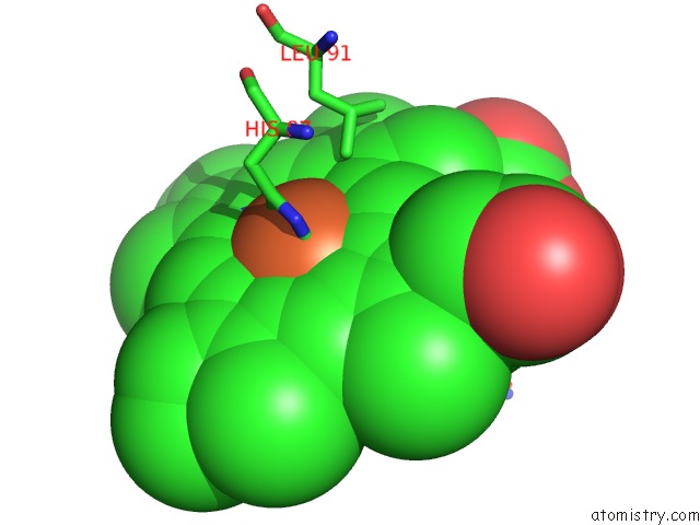
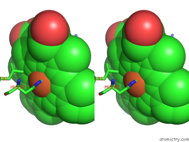
Iron binding site 1 out of 4 in 1a3o
Go back to
Iron binding site 1 out
of 4 in the Artificial Mutant (Alpha Y42H) of Deoxy Hemoglobin
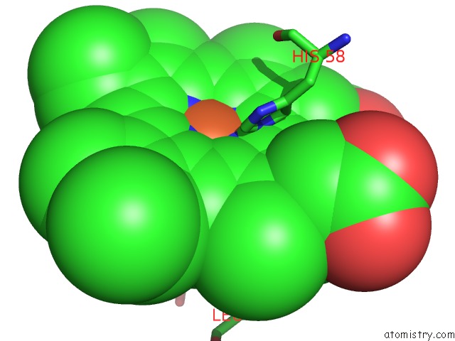
Mono view
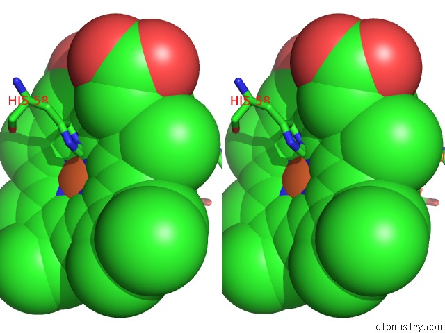
Stereo pair view
Mono view
Stereo pair view
A full contact list of Iron with other atoms in the Fe binding
site number 1 of Artificial Mutant (Alpha Y42H) of Deoxy Hemoglobin within 5.0Å range:
|
Iron binding site 2 out of 4 in 1a3o
Go back to
Iron binding site 2 out
of 4 in the Artificial Mutant (Alpha Y42H) of Deoxy Hemoglobin
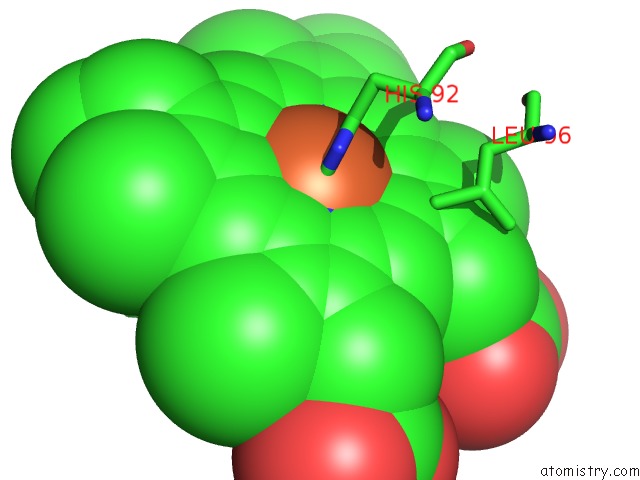
Mono view
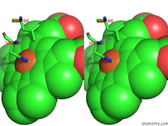
Stereo pair view
Mono view
Stereo pair view
A full contact list of Iron with other atoms in the Fe binding
site number 2 of Artificial Mutant (Alpha Y42H) of Deoxy Hemoglobin within 5.0Å range:
|
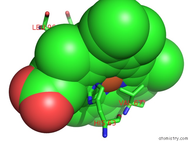
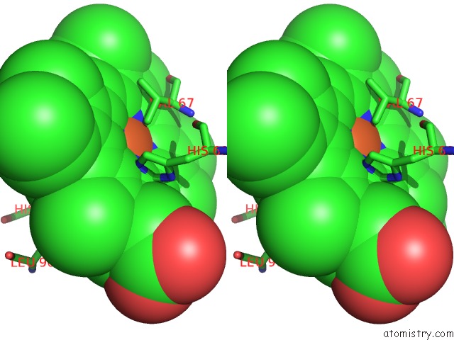
Iron binding site 3 out of 4 in 1a3o
Go back to
Iron binding site 3 out
of 4 in the Artificial Mutant (Alpha Y42H) of Deoxy Hemoglobin
Mono view
Stereo pair view
Mono view
Stereo pair view
A full contact list of Iron with other atoms in the Fe binding
site number 3 of Artificial Mutant (Alpha Y42H) of Deoxy Hemoglobin within 5.0Å range:
|
Iron binding site 4 out of 4 in 1a3o
Go back to
Iron binding site 4 out
of 4 in the Artificial Mutant (Alpha Y42H) of Deoxy Hemoglobin
Mono view
Stereo pair view
Mono view
Stereo pair view
A full contact list of Iron with other atoms in the Fe binding
site number 4 of Artificial Mutant (Alpha Y42H) of Deoxy Hemoglobin within 5.0Å range:
|
Reference:
J.R.Tame,
B.Vallone.
The Structures of Deoxy Human Haemoglobin and the Mutant Hb TYRALPHA42HIS at 120 K. Acta Crystallogr.,Sect.D V. 56 805 2000.
ISSN: ISSN 0907-4449
PubMed: 10930827
DOI: 10.1107/S0907444900006387
Page generated: Wed Jul 16 12:01:20 2025
ISSN: ISSN 0907-4449
PubMed: 10930827
DOI: 10.1107/S0907444900006387
Last articles
Fe in 2YXOFe in 2YRS
Fe in 2YXC
Fe in 2YNM
Fe in 2YVJ
Fe in 2YP1
Fe in 2YU2
Fe in 2YU1
Fe in 2YQB
Fe in 2YOO